Department of Cardiothoracic Surgery, Xinhua Hospital Affiliated to Shanghai Jiao Tong University School of Medicine, 1665 Kongjiang Road, Yangpu District, Shanghai, China.
Department of Pediatric Cardiovascular Surgery, Xinhua Hospital Affiliated to Shanghai Jiao Tong University School of Medicine, Shanghai, China.
Cell Biol Toxicol. 2024 May 27;40(1):40. doi: 10.1007/s10565-024-09873-6.
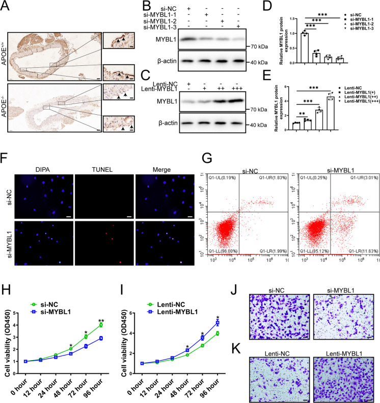
MYBL1 is a strong transcriptional activator involved in the cell signaling. However, there is no systematic study on the role of MYBL1 in atherosclerosis. The aim of this study is to elucidate the role and mechanism of MYBL1 in atherosclerosis. GSE28829, GSE43292 and GSE41571 were downloaded from NCBI for differentially expressed analysis. The expression levels of MYBL1 in atherosclerotic plaque tissue and normal vessels were detected by qRT-PCR, Western blot and Immunohistochemistry. Transwell and CCK-8 were used to detect the migration and proliferation of HUVECs after silencing MYBL1. RNA-seq, Western blot, qRT-PCR, Luciferase reporter system, Immunofluorescence, Flow cytometry, ChIP and CO-IP were used to study the role and mechanism of MYBL1 in atherosclerosis. The microarray data of GSE28829, GSE43292, and GSE41571 were analyzed and intersected, and then MYBL1 were verified. MYBL1 was down-regulated in atherosclerotic plaque tissue. After silencing of MYBL1, HUVECs were damaged, and their migration and proliferation abilities were weakened. Overexpression of MYBL1 significantly enhanced the migration and proliferation of HUVECs. MYBL1 knockdown induced abnormal autophagy in HUVEC cells, suggesting that MYBL1 was involved in the regulation of HUVECs through autophagy. Mechanistic studies showed that MYBL1 knockdown inhibited autophagosome and lysosomal fusion in HUVECs by inhibiting PLEKHM1, thereby exacerbating atherosclerosis. Furthermore, MYBL1 was found to repress lipid accumulation in HUVECs after oxLDL treatment. MYBL1 knockdown in HUVECs was involved in atherosclerosis by inhibiting PLEKHM1-induced autophagy, which provided a novel target of therapy for atherosclerosis.
MYBL1 是一种参与细胞信号转导的强转录激活因子。然而,目前尚没有关于 MYBL1 在动脉粥样硬化中的作用的系统研究。本研究旨在阐明 MYBL1 在动脉粥样硬化中的作用和机制。从 NCBI 下载 GSE28829、GSE43292 和 GSE41571 进行差异表达分析。通过 qRT-PCR、Western blot 和免疫组织化学检测动脉粥样硬化斑块组织和正常血管中 MYBL1 的表达水平。使用 Transwell 和 CCK-8 检测沉默 MYBL1 后 HUVECs 的迁移和增殖。RNA-seq、Western blot、qRT-PCR、荧光素酶报告系统、免疫荧光、流式细胞术、ChIP 和 CO-IP 用于研究 MYBL1 在动脉粥样硬化中的作用和机制。分析并交叉 GSE28829、GSE43292 和 GSE41571 的微阵列数据,然后验证 MYBL1。MYBL1 在动脉粥样硬化斑块组织中下调。沉默 MYBL1 后,HUVEC 受损,迁移和增殖能力减弱。过表达 MYBL1 可显著增强 HUVEC 的迁移和增殖能力。沉默 MYBL1 可诱导 HUVEC 细胞中异常自噬,表明 MYBL1 通过自噬参与 HUVEC 调节。机制研究表明,沉默 MYBL1 通过抑制 PLEKHM1 抑制 HUVEC 中的自噬体和溶酶体融合,从而加重动脉粥样硬化。此外,还发现 MYBL1 可抑制 oxLDL 处理后的 HUVEC 中的脂质积累。沉默 HUVEC 中的 MYBL1 通过抑制 PLEKHM1 诱导的自噬参与动脉粥样硬化,为动脉粥样硬化的治疗提供了新的靶点。