Boschen Suelen L, Seethaler Julian, Wang Shaohua, Lujan Wendy D, Silvernail Jodi L, Carter Rickey E, Chang Su-Youne, Lujan J Luis
Mayo Clinic.
Paracelsus Medical University.
Res Sq. 2024 May 14:rs.3.rs-4365911. doi: 10.21203/rs.3.rs-4365911/v1.
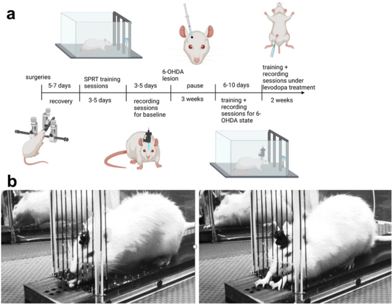
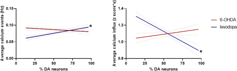
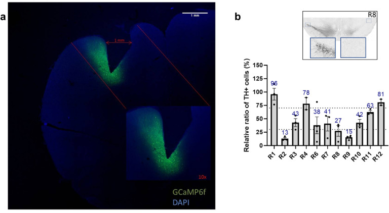
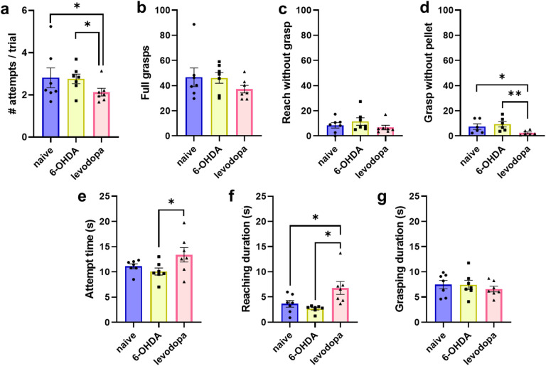
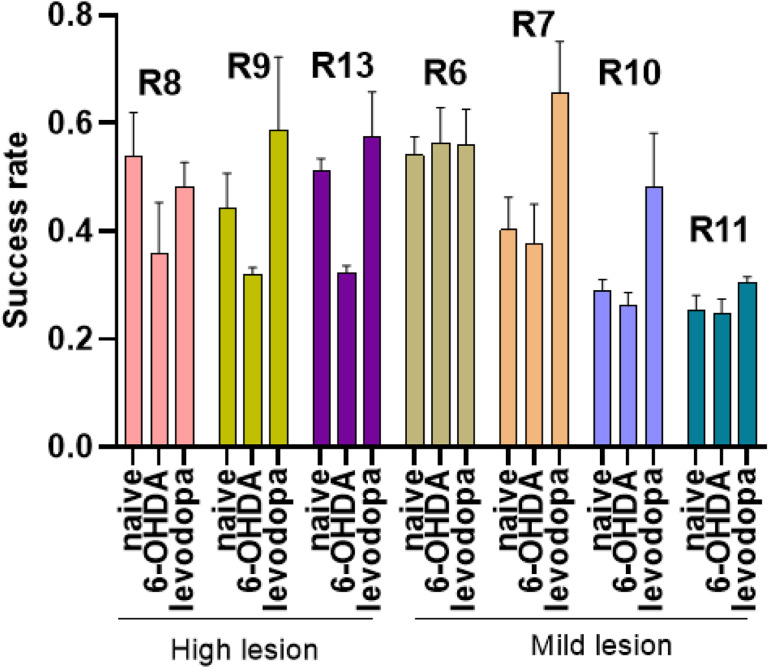
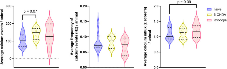
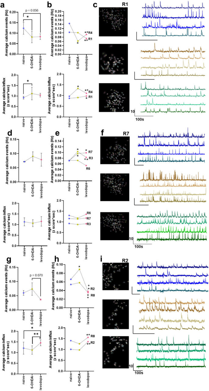
Parkinson's disease (PD) is marked by degeneration in the nigrostriatal dopaminergic pathway, affecting motor control via complex changes in the cortico-basal ganglia-thalamic motor network, including the primary motor cortex (M1). The modulation of M1 neuronal activity by dopaminergic inputs, particularly from the ventral tegmental area (VTA) and substantia nigra pars compacta (SNc), plays a crucial role in PD pathophysiology. This study investigates how nigrostriatal dopaminergic degeneration influences M1 neuronal activity in rats using in vivo calcium imaging. Histological analysis confirmed dopaminergic lesion severity, with high lesion level rats showing significant motor deficits. Levodopa treatment improved fine motor abilities, particularly in high lesion level rats. Analysis of M1 calcium signals based on dopaminergic lesion severity revealed distinct M1 activity patterns. Animals with low dopaminergic lesion showed increased calcium events, while high lesion level rats exhibited decreased activity, partially restored by levodopa. These findings suggest that M1 activity is more sensitive to transient fluctuations in dopaminergic transmission, rather than to chronic high or low dopaminergic signaling. This study underscores the complex interplay between dopaminergic signaling and M1 neuronal activity in PD symptoms development. Further research integrating behavioral and calcium imaging data can elucidate mechanisms underlying motor deficits and therapeutic responses in PD.
帕金森病(PD)的特征是黑质纹状体多巴胺能通路退化,通过皮质-基底神经节-丘脑运动网络(包括初级运动皮层(M1))的复杂变化影响运动控制。多巴胺能输入,特别是来自腹侧被盖区(VTA)和黑质致密部(SNc)的输入对M1神经元活动的调节在PD病理生理学中起着关键作用。本研究使用体内钙成像研究黑质纹状体多巴胺能退化如何影响大鼠的M1神经元活动。组织学分析证实了多巴胺能损伤的严重程度,高损伤水平的大鼠表现出明显的运动缺陷。左旋多巴治疗改善了精细运动能力,尤其是在高损伤水平的大鼠中。基于多巴胺能损伤严重程度对M1钙信号进行分析,揭示了不同的M1活动模式。多巴胺能损伤低的动物钙事件增加,而高损伤水平的大鼠活动减少,左旋多巴部分恢复了这种活动。这些发现表明,M1活动对多巴胺能传递的短暂波动更敏感,而不是对慢性高或低多巴胺能信号敏感。本研究强调了多巴胺能信号与M1神经元活动在PD症状发展中的复杂相互作用。进一步整合行为和钙成像数据的研究可以阐明PD运动缺陷和治疗反应的潜在机制。