Department of Clinical Medicine, Jining Medical University, Jining, China.
Department of Emergency Stroke, Affiliated Hospital of Jining Medical University, Jining, China.
J Neuroinflammation. 2024 May 28;21(1):140. doi: 10.1186/s12974-024-03113-8.
Perihematomal edema (PHE) after post-intracerebral hemorrhage (ICH) has complex pathophysiological mechanisms that are poorly understood. The complicated immune response in the post-ICH brain constitutes a crucial component of PHE pathophysiology. In this study, we aimed to characterize the transcriptional profiles of immune cell populations in human PHE tissue and explore the microscopic differences between different types of immune cells.
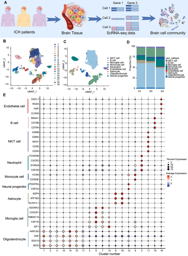
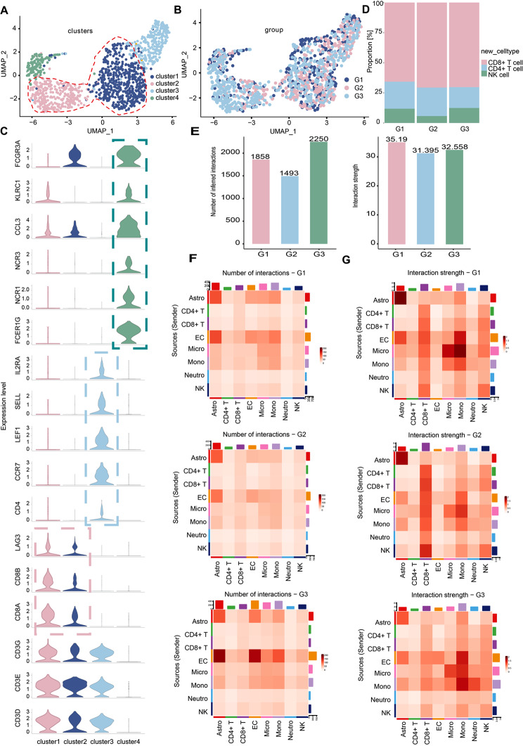
9 patients with basal ganglia intracerebral hemorrhage (hematoma volume 50-100 ml) were enrolled in this study. A multi-stage profile was developed, comprising Group1 (n = 3, 0-6 h post-ICH, G1), Group2 (n = 3, 6-24 h post-ICH, G2), and Group3 (n = 3, 24-48 h post-ICH, G3). A minimal quantity of edematous tissue surrounding the hematoma was preserved during hematoma evacuation. Single cell RNA sequencing (scRNA-seq) was used to map immune cell populations within comprehensively resected PHE samples collected from patients at different stages after ICH.
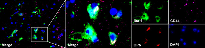
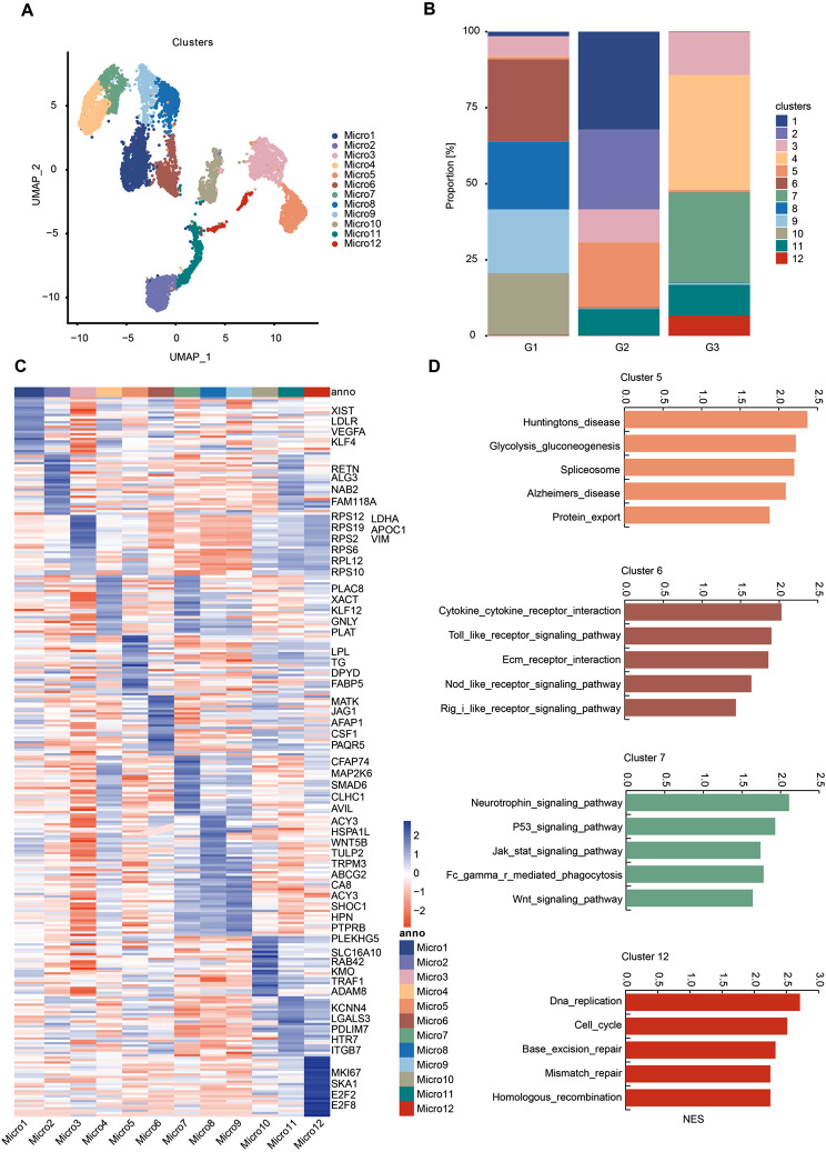
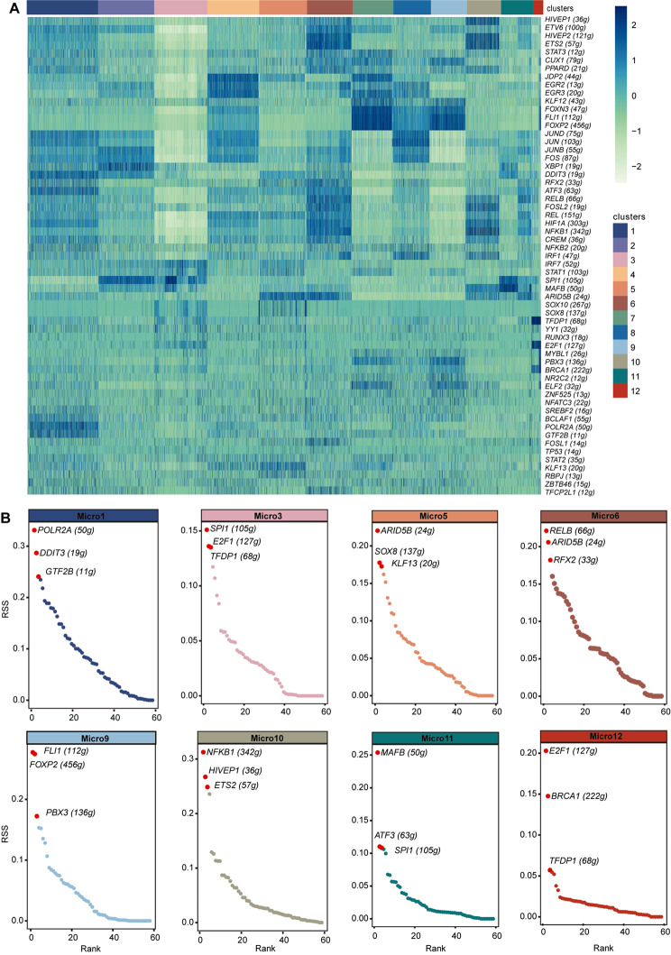
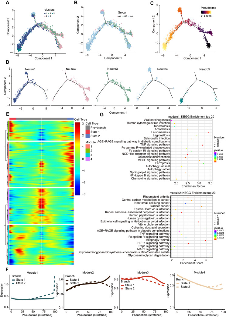
We established, for the first time, a comprehensive landscape of diverse immune cell populations in human PHE tissue at a single-cell level. Our study identified 12 microglia subsets and 5 neutrophil subsets in human PHE tissue. What's more, we discovered that the secreted phosphoprotein-1 (SPP1) pathway served as the basis for self-communication between microglia subclusters during the progression of PHE. Additionally, we traced the trajectory branches of different neutrophil subtypes. Finally, we also demonstrated that microglia-produced osteopontin (OPN) could regulate the immune environment in PHE tissue by interacting with CD44-positive cells.
As a result of our research, we have gained valuable insight into the immune-microenvironment within PHE tissue, which could potentially be used to develop novel treatment modalities for ICH.
脑出血(ICH)后出现的血肿周围水肿(PHE)具有复杂的病理生理学机制,目前尚未完全阐明。ICH 后脑内复杂的免疫反应是 PHE 病理生理学的重要组成部分。在这项研究中,我们旨在描述人类 PHE 组织中免疫细胞群体的转录谱,并探索不同类型免疫细胞之间的微观差异。
本研究纳入了 9 名基底节脑出血(血肿量 50-100ml)患者。采用多阶段方案,包括第 1 组(n=3,ICH 后 0-6 小时,G1)、第 2 组(n=3,ICH 后 6-24 小时,G2)和第 3 组(n=3,ICH 后 24-48 小时,G3)。在血肿清除过程中,保留了血肿周围少量水肿组织。使用单细胞 RNA 测序(scRNA-seq)对从不同 ICH 后阶段的患者中全面切除的 PHE 样本中免疫细胞群体进行了图谱绘制。
我们首次在单细胞水平上建立了人类 PHE 组织中多种免疫细胞群体的全面图谱。我们的研究在人类 PHE 组织中鉴定出 12 个小胶质细胞亚群和 5 个中性粒细胞亚群。此外,我们发现 SPP1 途径是 PHE 进展中小胶质细胞亚群之间自我通讯的基础。另外,我们追踪了不同中性粒细胞亚型的轨迹分支。最后,我们还证明了小胶质细胞产生的骨桥蛋白(OPN)可以通过与 CD44 阳性细胞相互作用来调节 PHE 组织中的免疫环境。
通过我们的研究,我们深入了解了 PHE 组织中的免疫微环境,这可能为 ICH 的治疗提供新的方法。