• 文献检索
  • 文档翻译
  • 深度研究
  • 学术资讯
  • Suppr Zotero 插件Zotero 插件
  • 邀请有礼
  • 套餐&价格
  • 历史记录
应用&插件
Suppr Zotero 插件Zotero 插件浏览器插件Mac 客户端Windows 客户端微信小程序
定价
高级版会员购买积分包购买API积分包
服务
文献检索文档翻译深度研究API 文档MCP 服务
关于我们
关于 Suppr公司介绍联系我们用户协议隐私条款
关注我们

Suppr 超能文献

核心技术专利:CN118964589B侵权必究
粤ICP备2023148730 号-1Suppr @ 2026

文献检索

告别复杂PubMed语法,用中文像聊天一样搜索,搜遍4000万医学文献。AI智能推荐,让科研检索更轻松。

立即免费搜索

文件翻译

保留排版,准确专业,支持PDF/Word/PPT等文件格式,支持 12+语言互译。

免费翻译文档

深度研究

AI帮你快速写综述,25分钟生成高质量综述,智能提取关键信息,辅助科研写作。

立即免费体验

在多扫描仪、多模态、质量各异的磁共振成像中对内侧颞叶亚区域进行自动分割。

Automatic segmentation of medial temporal lobe subregions in multi-scanner, multi-modality MRI of variable quality.

作者信息

Li Yue, Xie Long, Khandelwal Pulkit, Wisse Laura E M, Brown Christopher A, Prabhakaran Karthik, Dylan Tisdall M, Mechanic-Hamilton Dawn, Detre John A, Das Sandhitsu R, Wolk David A, Yushkevich Paul A

机构信息

Penn Image Computing and Science Laboratory, University of Pennsylvania, Philadelphia, USA.

Department of Radiology, University of Pennsylvania, Philadelphia, USA.

出版信息

bioRxiv. 2024 May 23:2024.05.21.595190. doi: 10.1101/2024.05.21.595190.

DOI:10.1101/2024.05.21.595190
PMID:38826413
原文链接:https://pmc.ncbi.nlm.nih.gov/articles/PMC11142184/
Abstract

BACKGROUND

Volumetry of subregions in the medial temporal lobe (MTL) computed from automatic segmentation in MRI can track neurodegeneration in Alzheimer's disease. However, image quality may vary in MRI. Poor quality MR images can lead to unreliable segmentation of MTL subregions. Considering that different MRI contrast mechanisms and field strengths (jointly referred to as "modalities" here) offer distinct advantages in imaging different parts of the MTL, we developed a muti-modality segmentation model using both 7 tesla (7T) and 3 tesla (3T) structural MRI to obtain robust segmentation in poor-quality images.

METHOD

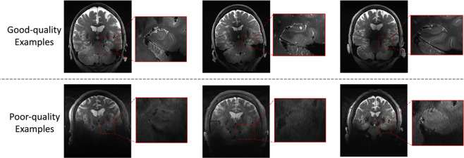
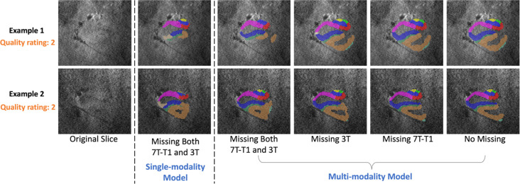
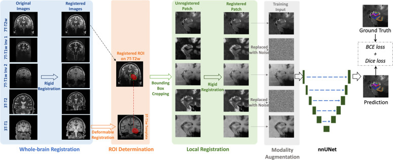
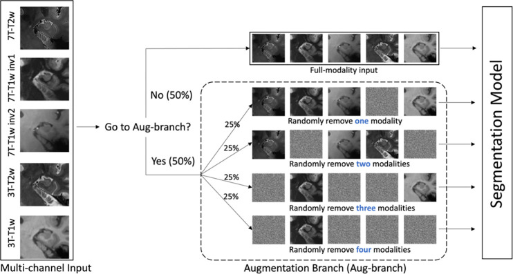
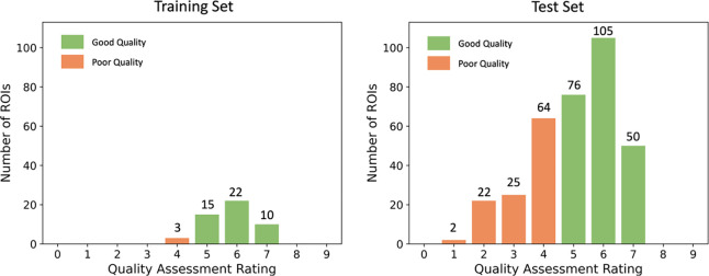
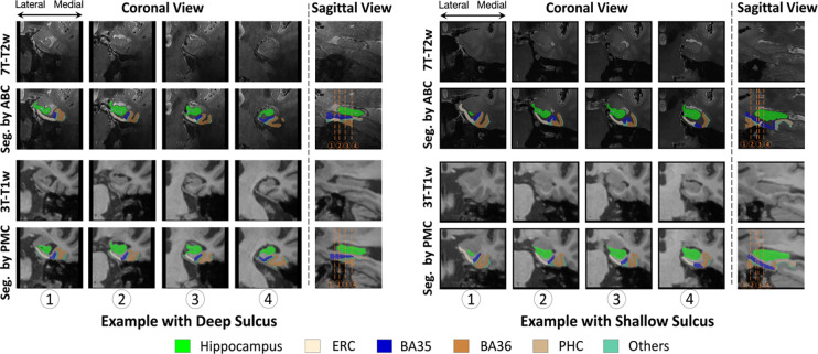
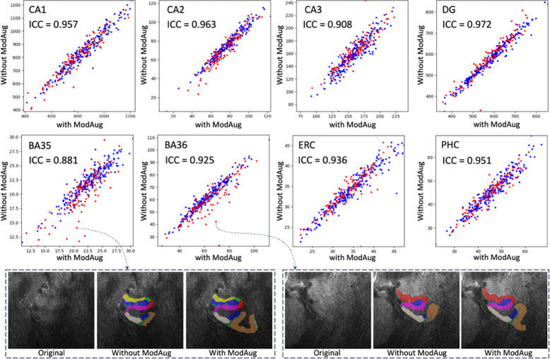
MRI modalities including 3T T1-weighted, 3T T2-weighted, 7T T1-weighted and 7T T2-weighted (7T-T2w) of 197 participants were collected from a longitudinal aging study at the Penn Alzheimer's Disease Research Center. Among them, 7T-T2w was used as the primary modality, and all other modalities were rigidly registered to the 7T-T2w. A model derived from nnU-Net took these registered modalities as input and outputted subregion segmentation in 7T-T2w space. 7T-T2w images most of which had high quality from 25 selected training participants were manually segmented to train the multi-modality model. Modality augmentation, which randomly replaced certain modalities with Gaussian noise, was applied during training to guide the model to extract information from all modalities. To compare our proposed model with a baseline single-modality model in the full dataset with mixed high/poor image quality, we evaluated the ability of derived volume/thickness measures to discriminate Amyloid+ mild cognitive impairment (A+MCI) and Amyloid- cognitively unimpaired (A-CU) groups, as well as the stability of these measurements in longitudinal data.

RESULTS

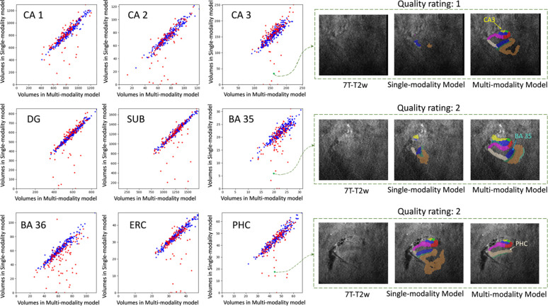
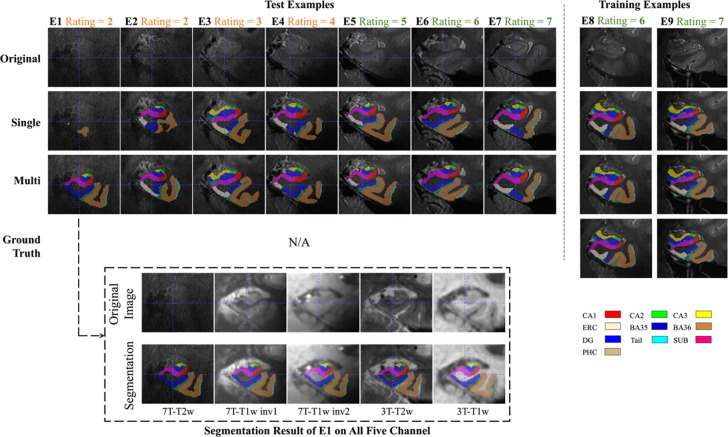
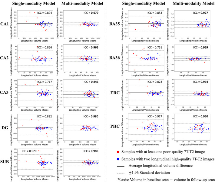
The multi-modality model delivered good performance regardless of 7T-T2w quality, while the single-modality model under-segmented subregions in poor-quality images. The multi-modality model generally demonstrated stronger discrimination of A+MCI versus A-CU. Intra-class correlation and Bland-Altman plots demonstrate that the multi-modality model had higher longitudinal segmentation consistency in all subregions while the single-modality model had low consistency in poor-quality images.

CONCLUSION

The multi-modality MRI segmentation model provides an improved biomarker for neurodegeneration in the MTL that is robust to image quality. It also provides a framework for other studies which may benefit from multimodal imaging.

摘要

背景

通过磁共振成像(MRI)自动分割计算得出的内侧颞叶(MTL)各亚区域体积,可追踪阿尔茨海默病中的神经退行性变。然而,MRI图像质量可能存在差异。质量较差的MR图像会导致MTL亚区域分割不可靠。鉴于不同的MRI对比机制和场强(在此统称为“模态”)在成像MTL不同部位时具有不同优势,我们开发了一种多模态分割模型,使用7特斯拉(7T)和3特斯拉(3T)结构MRI来在质量较差的图像中获得可靠的分割结果。

方法

从宾夕法尼亚阿尔茨海默病研究中心的一项纵向衰老研究中收集了197名参与者的MRI模态数据,包括3T T1加权、3T T2加权、7T T1加权和7T T2加权(7T-T2w)图像。其中,7T-T2w被用作主要模态,所有其他模态都被刚性配准到7T-T2w。一个源自nnU-Net的模型将这些配准后的模态作为输入,并输出7T-T2w空间中的亚区域分割结果。从25名选定的训练参与者中选取了大部分高质量的7T-T2w图像进行手动分割,以训练多模态模型。在训练过程中应用了模态增强,即随机用高斯噪声替换某些模态,以引导模型从所有模态中提取信息。为了在具有混合高/低图像质量的完整数据集中将我们提出的模型与基线单模态模型进行比较,我们评估了导出的体积/厚度测量值区分淀粉样蛋白阳性轻度认知障碍(A+MCI)和淀粉样蛋白阴性认知未受损(A-CU)组的能力,以及这些测量值在纵向数据中的稳定性。

结果

无论7T-T2w质量如何,多模态模型都表现出良好的性能,而单模态模型在质量较差的图像中对亚区域的分割不足。多模态模型通常对A+MCI与A-CU的区分能力更强。类内相关性和布兰德-奥特曼图表明,多模态模型在所有亚区域中具有更高的纵向分割一致性,而单模态模型在质量较差的图像中一致性较低。

结论

多模态MRI分割模型为MTL中的神经退行性变提供了一种改进的生物标志物,对图像质量具有鲁棒性。它还为其他可能受益于多模态成像的研究提供了一个框架。

https://cdn.ncbi.nlm.nih.gov/pmc/blobs/4522/11142184/c9ae9cd7ce82/nihpp-2024.05.21.595190v1-f0011.jpg
https://cdn.ncbi.nlm.nih.gov/pmc/blobs/4522/11142184/afd83567adbc/nihpp-2024.05.21.595190v1-f0001.jpg
https://cdn.ncbi.nlm.nih.gov/pmc/blobs/4522/11142184/53aacc4961bf/nihpp-2024.05.21.595190v1-f0002.jpg
https://cdn.ncbi.nlm.nih.gov/pmc/blobs/4522/11142184/03417cc324c6/nihpp-2024.05.21.595190v1-f0003.jpg
https://cdn.ncbi.nlm.nih.gov/pmc/blobs/4522/11142184/870969b7d05f/nihpp-2024.05.21.595190v1-f0004.jpg
https://cdn.ncbi.nlm.nih.gov/pmc/blobs/4522/11142184/d3b88571b5f8/nihpp-2024.05.21.595190v1-f0005.jpg
https://cdn.ncbi.nlm.nih.gov/pmc/blobs/4522/11142184/ab6a3156051a/nihpp-2024.05.21.595190v1-f0006.jpg
https://cdn.ncbi.nlm.nih.gov/pmc/blobs/4522/11142184/7a9536960161/nihpp-2024.05.21.595190v1-f0007.jpg
https://cdn.ncbi.nlm.nih.gov/pmc/blobs/4522/11142184/1981a3837af5/nihpp-2024.05.21.595190v1-f0008.jpg
https://cdn.ncbi.nlm.nih.gov/pmc/blobs/4522/11142184/3887cf453d78/nihpp-2024.05.21.595190v1-f0009.jpg
https://cdn.ncbi.nlm.nih.gov/pmc/blobs/4522/11142184/a741b7e67180/nihpp-2024.05.21.595190v1-f0010.jpg
https://cdn.ncbi.nlm.nih.gov/pmc/blobs/4522/11142184/c9ae9cd7ce82/nihpp-2024.05.21.595190v1-f0011.jpg

相似文献

1
Automatic segmentation of medial temporal lobe subregions in multi-scanner, multi-modality MRI of variable quality.在多扫描仪、多模态、质量各异的磁共振成像中对内侧颞叶亚区域进行自动分割。
bioRxiv. 2024 May 23:2024.05.21.595190. doi: 10.1101/2024.05.21.595190.
2
Clinical Application of Automatic Segmentation of Medial Temporal Lobe Subregions in Prodromal and Dementia-Level Alzheimer's Disease.内侧颞叶亚区域自动分割在前驱期和痴呆级阿尔茨海默病中的临床应用
J Alzheimers Dis. 2016 Oct 4;54(3):1027-1037. doi: 10.3233/JAD-160014.
3
Automatic Hippocampal Subfield Segmentation from 3T Multi-modality Images.基于3T多模态图像的海马亚区自动分割
Mach Learn Med Imaging. 2016 Oct;10019:229-236. doi: 10.1007/978-3-319-47157-0_28. Epub 2016 Oct 1.
4
Medial Temporal Lobe Subregional Atrophy in Aging and Alzheimer's Disease: A Longitudinal Study.衰老和阿尔茨海默病中的内侧颞叶亚区域萎缩:一项纵向研究。
Front Aging Neurosci. 2021 Oct 15;13:750154. doi: 10.3389/fnagi.2021.750154. eCollection 2021.
5
Morphometry of medial temporal lobe subregions using high-resolution T2-weighted MRI in ADNI3: Why, how, and what's next?ADNI3 中使用高分辨率 T2 加权 MRI 测量内侧颞叶亚区的形态计量学:为什么、如何以及下一步是什么?
Alzheimers Dement. 2024 Nov;20(11):8113-8128. doi: 10.1002/alz.14161. Epub 2024 Sep 16.
6
Automated segmentation of medial temporal lobe subregions on in vivo T1-weighted MRI in early stages of Alzheimer's disease.阿尔茨海默病早期活体 T1 加权 MRI 上内侧颞叶亚区的自动分割。
Hum Brain Mapp. 2019 Aug 15;40(12):3431-3451. doi: 10.1002/hbm.24607. Epub 2019 Apr 29.
7
Automated Multi-Atlas Segmentation of Hippocampal and Extrahippocampal Subregions in Alzheimer's Disease at 3T and 7T: What Atlas Composition Works Best?在 3T 和 7T 下阿尔茨海默病的海马和海马外亚区的自动多图谱分割:哪种图谱组合效果最好?
J Alzheimers Dis. 2018;63(1):217-225. doi: 10.3233/JAD-170932.
8
Reconstruction of 7T-Like Images From 3T MRI.从3T磁共振成像重建类似7T的图像。
IEEE Trans Med Imaging. 2016 Sep;35(9):2085-97. doi: 10.1109/TMI.2016.2549918. Epub 2016 Apr 1.
9
Structural and functional asymmetry of medial temporal subregions in unilateral temporal lobe epilepsy: A 7T MRI study.单侧颞叶癫痫患者内侧颞叶亚区的结构和功能不对称:7T MRI 研究。
Hum Brain Mapp. 2019 Jun 1;40(8):2390-2398. doi: 10.1002/hbm.24530. Epub 2019 Jan 21.
10
Mapping the structural and functional network architecture of the medial temporal lobe using 7T MRI.使用 7T MRI 绘制内侧颞叶的结构和功能网络架构。
Hum Brain Mapp. 2018 Feb;39(2):851-865. doi: 10.1002/hbm.23887. Epub 2017 Nov 20.

本文引用的文献

1
Deep label fusion: A generalizable hybrid multi-atlas and deep convolutional neural network for medical image segmentation.深度标签融合:一种可推广的混合多图谱和深度卷积神经网络的医学图像分割方法。
Med Image Anal. 2023 Jan;83:102683. doi: 10.1016/j.media.2022.102683. Epub 2022 Nov 5.
2
Causal links among amyloid, tau, and neurodegeneration.淀粉样蛋白、tau蛋白与神经退行性变之间的因果联系。
Brain Commun. 2022 Jul 25;4(4):fcac193. doi: 10.1093/braincomms/fcac193. eCollection 2022.
3
Accurate and Robust Alignment of Differently Stained Histologic Images Based on Greedy Diffeomorphic Registration.
基于贪婪微分同胚配准的不同染色组织学图像的精确且稳健配准
Appl Sci (Basel). 2021 Feb;11(4). doi: 10.3390/app11041892. Epub 2021 Feb 21.
4
nnU-Net: a self-configuring method for deep learning-based biomedical image segmentation.nnU-Net:一种基于深度学习的生物医学图像分割的自配置方法。
Nat Methods. 2021 Feb;18(2):203-211. doi: 10.1038/s41592-020-01008-z. Epub 2020 Dec 7.
5
Hippocampal subfield volumetry from structural isotropic 1 mm MRI scans: A note of caution.结构各向同性 1mmMRI 扫描的海马亚区容积测量:注意事项。
Hum Brain Mapp. 2021 Feb 1;42(2):539-550. doi: 10.1002/hbm.25234. Epub 2020 Oct 15.
6
Longitudinal atrophy in early Braak regions in preclinical Alzheimer's disease.早期 Braak 阶段临床前阿尔茨海默病中的纵向萎缩。
Hum Brain Mapp. 2020 Nov;41(16):4704-4717. doi: 10.1002/hbm.25151. Epub 2020 Aug 26.
7
Association of Factors With Elevated Amyloid Burden in Clinically Normal Older Individuals.与临床正常老年人淀粉样蛋白负担增加相关的因素。
JAMA Neurol. 2020 Jun 1;77(6):735-745. doi: 10.1001/jamaneurol.2020.0387.
8
Association between subfield volumes of the medial temporal lobe and cognitive assessments.内侧颞叶亚区体积与认知评估之间的关联。
Heliyon. 2019 Jun 4;5(6):e01828. doi: 10.1016/j.heliyon.2019.e01828. eCollection 2019 Jun.
9
Automated Volumetry of Medial Temporal Lobe Subregions in Mild Cognitive Impairment and Alzheimer Disease.轻度认知障碍和阿尔茨海默病内侧颞叶亚区的自动容积测量。
Alzheimer Dis Assoc Disord. 2019 Jul-Sep;33(3):206-211. doi: 10.1097/WAD.0000000000000318.
10
Automated segmentation of medial temporal lobe subregions on in vivo T1-weighted MRI in early stages of Alzheimer's disease.阿尔茨海默病早期活体 T1 加权 MRI 上内侧颞叶亚区的自动分割。
Hum Brain Mapp. 2019 Aug 15;40(12):3431-3451. doi: 10.1002/hbm.24607. Epub 2019 Apr 29.