Xie An, Sun Yunkai, Chen Haobo, Li Ling, Liu Peng, Liao Yanhui, Li Yonggang
Department of Radiology, The First Affiliated Hospital of Soochow University, Suzhou, Jiangsu, China.
Department of Radiology, The People's Hospital of Hunan Province (The First Affiliated Hospital of Hunan Normal University), Changsha, Hunan, China.
Front Psychiatry. 2024 May 16;15:1353103. doi: 10.3389/fpsyt.2024.1353103. eCollection 2024.
Insular subdivisions show distinct patterns of resting state functional connectivity with specific brain regions, each with different functional significance in chronic cigarette smokers. This study aimed to explore the altered dynamic functional connectivity (dFC) of distinct insular subdivisions in smokers.
Resting-state BOLD data of 31 smokers with nicotine dependence and 27 age-matched non-smokers were collected. Three bilateral insular regions of interest (dorsal, ventral, and posterior) were set as seeds for analyses. Sliding windows method was used to acquire the dFC metrics of different insular seeds. Support vector machine based on abnormal insular dFC was applied to classify smokers from non-smokers.
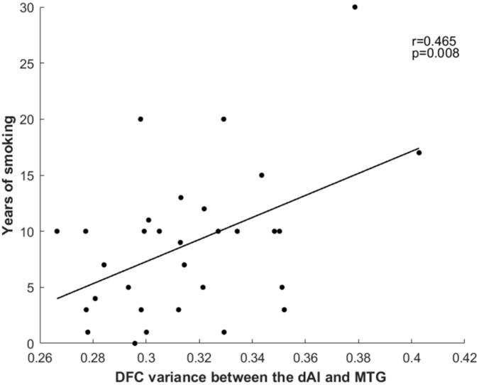
We found that smokers showed lower dFC variance between the left ventral anterior insula and both the right superior parietal cortex and the left inferior parietal cortex, as well as greater dFC variance the right ventral anterior insula with the right middle cingulum cortex relative to non-smokers. Moreover, compared to non-smokers, it is found that smokers demonstrated altered dFC variance of the right dorsal insula and the right middle temporal gyrus. Correlation analysis showed the higher dFC between the right dorsal insula and the right middle temporal gyrus was associated with longer years of smoking. The altered insular subdivision dFC can classify smokers from non-smokers with an accuracy of 89.66%, a sensitivity of 96.30% and a specify of 83.87%.
Our findings highlighted the abnormal patterns of fluctuating connectivity of insular subdivision circuits in smokers and suggested that these abnormalities may play a significant role in the mechanisms underlying nicotine addiction and could potentially serve as a neural biomarker for addiction treatment.
脑岛亚区与特定脑区呈现出不同的静息态功能连接模式,在慢性吸烟者中各自具有不同的功能意义。本研究旨在探究吸烟者不同脑岛亚区动态功能连接(dFC)的改变。
收集了31名尼古丁依赖吸烟者和27名年龄匹配的非吸烟者的静息态BOLD数据。将三个双侧脑岛感兴趣区(背侧、腹侧和后侧)设为分析种子点。采用滑动窗口法获取不同脑岛种子点的dFC指标。基于异常脑岛dFC的支持向量机用于区分吸烟者和非吸烟者。
我们发现,与非吸烟者相比,吸烟者左腹侧前脑岛与右侧顶上叶皮质和左侧顶下叶皮质之间的dFC方差较低,而右侧腹侧前脑岛与右侧中央扣带皮质之间的dFC方差较高。此外,与非吸烟者相比,吸烟者右侧背侧脑岛和右侧颞中回的dFC方差发生改变。相关性分析表明,右侧背侧脑岛与右侧颞中回之间较高的dFC与吸烟年限较长有关。脑岛亚区dFC的改变能够以89.66%的准确率、96.30%的灵敏度和83.87%的特异度区分吸烟者和非吸烟者。
我们的研究结果突出了吸烟者脑岛亚区回路波动连接的异常模式,并表明这些异常可能在尼古丁成瘾机制中起重要作用,并且有可能作为成瘾治疗的神经生物标志物。