• 文献检索
  • 文档翻译
  • 深度研究
  • 学术资讯
  • Suppr Zotero 插件Zotero 插件
  • 邀请有礼
  • 套餐&价格
  • 历史记录
应用&插件
Suppr Zotero 插件Zotero 插件浏览器插件Mac 客户端Windows 客户端微信小程序
定价
高级版会员购买积分包购买API积分包
服务
文献检索文档翻译深度研究API 文档MCP 服务
关于我们
关于 Suppr公司介绍联系我们用户协议隐私条款
关注我们

Suppr 超能文献

核心技术专利:CN118964589B侵权必究
粤ICP备2023148730 号-1Suppr @ 2026

文献检索

告别复杂PubMed语法,用中文像聊天一样搜索,搜遍4000万医学文献。AI智能推荐,让科研检索更轻松。

立即免费搜索

文件翻译

保留排版,准确专业,支持PDF/Word/PPT等文件格式,支持 12+语言互译。

免费翻译文档

深度研究

AI帮你快速写综述,25分钟生成高质量综述,智能提取关键信息,辅助科研写作。

立即免费体验

数字射线:使用风格迁移生成对抗网络增强白内障眼底图像,以提高视网膜病变检测。

Digital ray: enhancing cataractous fundus images using style transfer generative adversarial networks to improve retinopathy detection.

机构信息

Zhongshan Ophthalmic Center State Key Laboratory of Ophthalmology, Sun Yat-Sen University, Guangzhou, Guangdong, China.

School of Medical Information Engineering, Guangzhou University of Chinese Medicine, Guangzhou, China.

出版信息

Br J Ophthalmol. 2024 Sep 20;108(10):1423-1429. doi: 10.1136/bjo-2024-325403.

DOI:10.1136/bjo-2024-325403
PMID:38839251
原文链接:https://pmc.ncbi.nlm.nih.gov/articles/PMC11503040/
Abstract

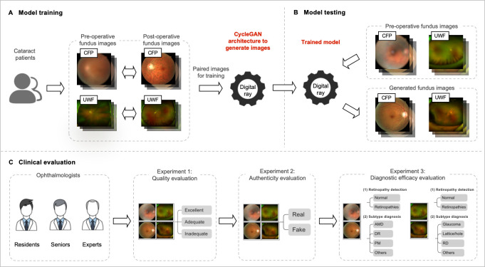
BACKGROUND/AIMS: The aim of this study was to develop and evaluate digital ray, based on preoperative and postoperative image pairs using style transfer generative adversarial networks (GANs), to enhance cataractous fundus images for improved retinopathy detection.

METHODS

For eligible cataract patients, preoperative and postoperative colour fundus photographs (CFP) and ultra-wide field (UWF) images were captured. Then, both the original CycleGAN and a modified CycleGAN (CycleGAN) framework were adopted for image generation and quantitatively compared using Frechet Inception Distance (FID) and Kernel Inception Distance (KID). Additionally, CFP and UWF images from another cataract cohort were used to test model performances. Different panels of ophthalmologists evaluated the quality, authenticity and diagnostic efficacy of the generated images.

RESULTS

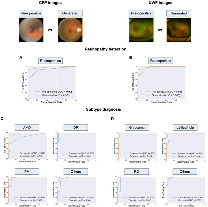
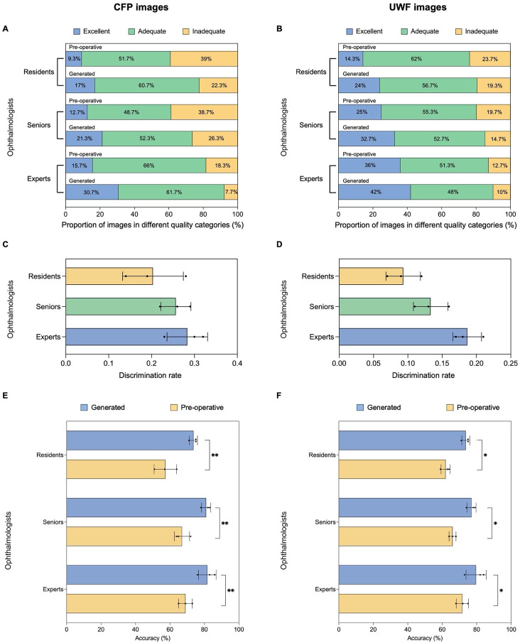
A total of 959 CFP and 1009 UWF image pairs were included in model development. FID and KID indicated that images generated by CycleGAN presented significantly improved quality. Based on ophthalmologists' average ratings, the percentages of inadequate-quality images decreased from 32% to 18.8% for CFP, and from 18.7% to 14.7% for UWF. Only 24.8% and 13.8% of generated CFP and UWF images could be recognised as synthetic. The accuracy of retinopathy detection significantly increased from 78% to 91% for CFP and from 91% to 93% for UWF. For retinopathy subtype diagnosis, the accuracies also increased from 87%-94% to 91%-100% for CFP and from 87%-95% to 93%-97% for UWF.

CONCLUSION

Digital ray could generate realistic postoperative CFP and UWF images with enhanced quality and accuracy for overall detection and subtype diagnosis of retinopathies, especially for CFP.\ TRIAL REGISTRATION NUMBER: This study was registered with ClinicalTrials.gov (NCT05491798).

摘要

背景/目的:本研究旨在开发一种基于术前和术后图像对的数字射线,使用风格转换生成对抗网络(GAN)增强白内障眼底图像,以提高糖尿病视网膜病变的检测效果。

方法

对符合条件的白内障患者进行术前和术后彩色眼底照相(CFP)和超广角(UWF)图像采集。然后,采用原始 CycleGAN 和修改后的 CycleGAN(CycleGAN)框架进行图像生成,并使用 Frechet Inception Distance(FID)和 Kernel Inception Distance(KID)进行定量比较。此外,还使用另一组白内障患者的 CFP 和 UWF 图像来测试模型性能。不同的眼科医生小组评估生成图像的质量、真实性和诊断效果。

结果

共纳入 959 对 CFP 和 1009 对 UWF 图像进行模型开发。FID 和 KID 表明,CycleGAN 生成的图像质量显著提高。根据眼科医生的平均评分,CFP 的低质量图像比例从 32%下降到 18.8%,UWF 的低质量图像比例从 18.7%下降到 14.7%。只有 24.8%和 13.8%的 CFP 和 UWF 生成图像被识别为合成图像。CFP 的糖尿病视网膜病变检测准确率从 78%提高到 91%,UWF 的准确率从 91%提高到 93%。对于糖尿病视网膜病变亚型诊断,准确率也从 CFP 的 87%-94%提高到 91%-100%,从 UWF 的 87%-95%提高到 93%-97%。

结论

数字射线可以生成逼真的术后 CFP 和 UWF 图像,提高图像质量和整体检测的准确性,有助于糖尿病视网膜病变及其亚型的诊断。

试验注册

本研究在 ClinicalTrials.gov 注册(NCT05491798)。

https://cdn.ncbi.nlm.nih.gov/pmc/blobs/2995/11503040/871d44ae5ddf/bjo-108-10-g003.jpg
https://cdn.ncbi.nlm.nih.gov/pmc/blobs/2995/11503040/a572dbb01713/bjo-108-10-g001.jpg
https://cdn.ncbi.nlm.nih.gov/pmc/blobs/2995/11503040/3532e3f72f95/bjo-108-10-g002.jpg
https://cdn.ncbi.nlm.nih.gov/pmc/blobs/2995/11503040/871d44ae5ddf/bjo-108-10-g003.jpg
https://cdn.ncbi.nlm.nih.gov/pmc/blobs/2995/11503040/a572dbb01713/bjo-108-10-g001.jpg
https://cdn.ncbi.nlm.nih.gov/pmc/blobs/2995/11503040/3532e3f72f95/bjo-108-10-g002.jpg
https://cdn.ncbi.nlm.nih.gov/pmc/blobs/2995/11503040/871d44ae5ddf/bjo-108-10-g003.jpg

相似文献

1
Digital ray: enhancing cataractous fundus images using style transfer generative adversarial networks to improve retinopathy detection.数字射线:使用风格迁移生成对抗网络增强白内障眼底图像,以提高视网膜病变检测。
Br J Ophthalmol. 2024 Sep 20;108(10):1423-1429. doi: 10.1136/bjo-2024-325403.
2
Deep learning-based classification of retinal vascular diseases using ultra-widefield colour fundus photographs.基于深度学习的超广角彩色眼底照片视网膜血管疾病分类。
BMJ Open Ophthalmol. 2022 Feb 4;7(1):e000924. doi: 10.1136/bmjophth-2021-000924. eCollection 2022.
3
ETDRS grading with CLARUS ultra-widefield images shows agreement with 7-fields colour fundus photography.ETDRS 分级与 CLARUS 超广角图像具有一致性,与 7 区域彩色眼底照相一致。
BMC Ophthalmol. 2024 Sep 3;24(1):387. doi: 10.1186/s12886-024-03537-z.
4
Comparison of widefield swept-source optical coherence tomography angiography with ultra-widefield colour fundus photography and fluorescein angiography for detection of lesions in diabetic retinopathy.宽视野扫频光学相干断层扫描血管造影与超广角彩色眼底照相和荧光素血管造影在糖尿病视网膜病变病变检测中的比较。
Br J Ophthalmol. 2021 Apr;105(4):577-581. doi: 10.1136/bjophthalmol-2020-316245. Epub 2020 Jun 26.
5
Ultra-widefield color fundus photography combined with high-speed ultra-widefield swept-source optical coherence tomography angiography for non-invasive detection of lesions in diabetic retinopathy.超广域彩色眼底照相联合高速超广域扫频源光相干断层扫描血管成像术无创检测糖尿病视网膜病变病变。
Front Public Health. 2022 Nov 2;10:1047608. doi: 10.3389/fpubh.2022.1047608. eCollection 2022.
6
Leveraging Regular Fundus Images for Training UWF Fundus Diagnosis Models via Adversarial Learning and Pseudo-Labeling.基于对抗学习和伪标记的常规眼底图像在超广角眼底诊断模型中的应用。
IEEE Trans Med Imaging. 2021 Oct;40(10):2911-2925. doi: 10.1109/TMI.2021.3056395. Epub 2021 Sep 30.
7
CycleGAN-based deep learning technique for artifact reduction in fundus photography.基于 CycleGAN 的深度学习技术在眼底摄影中减少伪影。
Graefes Arch Clin Exp Ophthalmol. 2020 Aug;258(8):1631-1637. doi: 10.1007/s00417-020-04709-5. Epub 2020 May 2.
8
ULTRAWIDEFIELD PSEUDOCOLOR RETINAL IMAGING VERSUS REAL-COLOR FUNDUS PHOTOGRAPHY FOR DETECTION OF INTRARETINAL PIGMENT MIGRATION IN AGE-RELATED MACULAR DEGENERATION.超宽视野伪彩视网膜成像与真彩眼底照相在年龄相关性黄斑变性中检测视网膜内色素迁移的比较。
Retina. 2021 Mar 1;41(3):563-571. doi: 10.1097/IAE.0000000000002886.
9
Investigations on whether image-sharpening algorithm can improve the determination of severity of diabetic retinopathy in ultra-widefield fundus photographs.关于图像锐化算法能否改善超广角眼底照片中糖尿病视网膜病变严重程度判定的研究。
Sci Rep. 2025 Apr 2;15(1):11351. doi: 10.1038/s41598-025-92270-8.
10
Prediction of Visual Acuity After Cataract Surgery by Deep Learning Methods Using Clinical Information and Color Fundus Photography.利用临床信息和彩色眼底照片,通过深度学习方法预测白内障手术后的视力
Curr Eye Res. 2025 Mar;50(3):276-281. doi: 10.1080/02713683.2024.2430212. Epub 2024 Dec 9.

引用本文的文献

1
Applications of generative adversarial networks in the diagnosis, prognosis, and treatment of ophthalmic diseases.生成对抗网络在眼科疾病诊断、预后及治疗中的应用。
Graefes Arch Clin Exp Ophthalmol. 2025 Apr 22. doi: 10.1007/s00417-025-06830-9.

本文引用的文献

1
DeepQuality improves infant retinopathy screening.深度质量改善婴儿视网膜病变筛查。
NPJ Digit Med. 2023 Oct 16;6(1):192. doi: 10.1038/s41746-023-00943-3.
2
DeepFundus: A flow-cytometry-like image quality classifier for boosting the whole life cycle of medical artificial intelligence.深眼底:一种类似流式细胞术的图像质量分类器,用于提升医疗人工智能的整个生命周期。
Cell Rep Med. 2023 Feb 21;4(2):100912. doi: 10.1016/j.xcrm.2022.100912. Epub 2023 Jan 19.
3
Real-time diabetic retinopathy screening by deep learning in a multisite national screening programme: a prospective interventional cohort study.
深度学习在多中心全国性筛查项目中实时筛查糖尿病视网膜病变:一项前瞻性干预性队列研究。
Lancet Digit Health. 2022 Apr;4(4):e235-e244. doi: 10.1016/S2589-7500(22)00017-6. Epub 2022 Mar 7.
4
An Annotation-Free Restoration Network for Cataractous Fundus Images.无注释白内障眼底图像恢复网络。
IEEE Trans Med Imaging. 2022 Jul;41(7):1699-1710. doi: 10.1109/TMI.2022.3147854. Epub 2022 Jun 30.
5
Image quality issues in teledermatology: A comparative analysis of artificial intelligence solutions.远程皮肤病学中的图像质量问题:人工智能解决方案的比较分析
J Am Acad Dermatol. 2022 Jul;87(1):240-242. doi: 10.1016/j.jaad.2021.07.073. Epub 2021 Aug 10.
6
Structure and Illumination Constrained GAN for Medical Image Enhancement.结构和光照约束生成对抗网络在医学图像增强中的应用。
IEEE Trans Med Imaging. 2021 Dec;40(12):3955-3967. doi: 10.1109/TMI.2021.3101937. Epub 2021 Nov 30.
7
The Clinician and Dataset Shift in Artificial Intelligence.临床医生与人工智能中的数据集偏移
N Engl J Med. 2021 Jul 15;385(3):283-286. doi: 10.1056/NEJMc2104626.
8
Management of Cataract in Patients with Age-Related Macular Degeneration.年龄相关性黄斑变性患者的白内障管理
J Clin Med. 2021 Jun 8;10(12):2538. doi: 10.3390/jcm10122538.
9
Modeling and Enhancing Low-Quality Retinal Fundus Images.眼底低质量图像的建模与增强。
IEEE Trans Med Imaging. 2021 Mar;40(3):996-1006. doi: 10.1109/TMI.2020.3043495. Epub 2021 Mar 2.
10
Deep learning from "passive feeding" to "selective eating" of real-world data.从对现实世界数据的“被动摄取”到“选择性取用”的深度学习。
NPJ Digit Med. 2020 Oct 30;3:143. doi: 10.1038/s41746-020-00350-y. eCollection 2020.