Institute of Scientific Instruments of the Czech Academy of Sciences, Královopolská 147, Brno, 612 00, Czech Republic.
Institute of Experimental Botany of the Czech Academy of Sciences, Centre of Plant Structural and Functional Genomics, Šlechtitelů 31, Olomouc, 772 00, Czech Republic.
Sci Rep. 2024 Jun 6;14(1):12998. doi: 10.1038/s41598-024-63515-9.
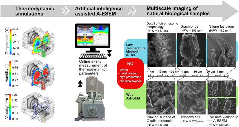
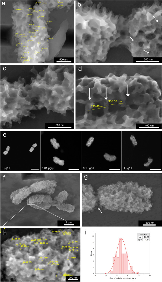
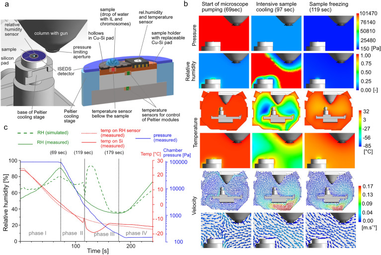
The challenge of in-situ handling and high-resolution low-dose imaging of intact, sensitive and wet samples in their native state at nanometer scale, including live samples is met by Advanced Environmental Scanning Electron Microscopy (A-ESEM). This new generation of ESEM utilises machine learning-based optimization of thermodynamic conditions with respect to sample specifics to employ a low temperature method and an ionization secondary electron detector with an electrostatic separator. A modified electron microscope was used, equipped with temperature, humidity and gas pressure sensors for in-situ and real-time monitoring of the sample. A transparent ultra-thin film of ionic liquid is used to increase thermal and electrical conductivity of the samples and to minimize sample damage by free radicals. To validate the power of the new method, we analyze condensed mitotic metaphase chromosomes to reveal new structural features of their perichromosomal layer, and the organization of chromatin fibers, not observed before by any microscopic technique. The ability to resolve nano-structural details of chromosomes using A-ESEM is validated by measuring gold nanoparticles with achievable resolution in the lower nanometre units.
原位处理和在纳米尺度上对完整、敏感和湿润的天然状态样品进行高分辨率低剂量成像的挑战,包括活样本,都可以通过先进环境扫描电子显微镜(A-ESEM)来解决。这种新一代的 ESEM 利用基于机器学习的热力学条件优化,针对样品的具体情况,采用低温方法和带有静电分离器的离子二次电子探测器。我们使用了经过改良的电子显微镜,配备了温度、湿度和气压传感器,用于对样品进行原位和实时监测。我们使用透明的超薄离子液体薄膜来提高样品的热导率和电导率,并最大程度地减少自由基对样品的损伤。为了验证新方法的有效性,我们分析了浓缩的有丝分裂中期染色体,以揭示其周边层的新结构特征,以及染色质纤维的组织,这是以前任何显微镜技术都没有观察到的。通过使用 A-ESEM 测量可达纳米以下分辨率的金纳米粒子,验证了其对染色体纳米结构细节的分辨能力。