Charité - Universitätsmedizin Berlin, corporate member of Freie Universität Berlin and Humboldt-Universität zu Berlin, Experimental and Clinical Research Center, Berlin, Germany.
Neuroscience Clinical Research Center, Charité - Universitätsmedizin Berlin, corporate member of Freie Universität Berlin and Humboldt-Universität zu Berlin, Berlin, Germany.
Ann Clin Transl Neurol. 2024 Aug;11(8):2016-2029. doi: 10.1002/acn3.52121. Epub 2024 Jun 14.
Persisting neurological symptoms after COVID-19 affect up to 10% of patients and can manifest in fatigue and cognitive complaints. Based on recent evidence, we evaluated whether cerebral hemodynamic changes contribute to post-COVID syndrome (PCS).
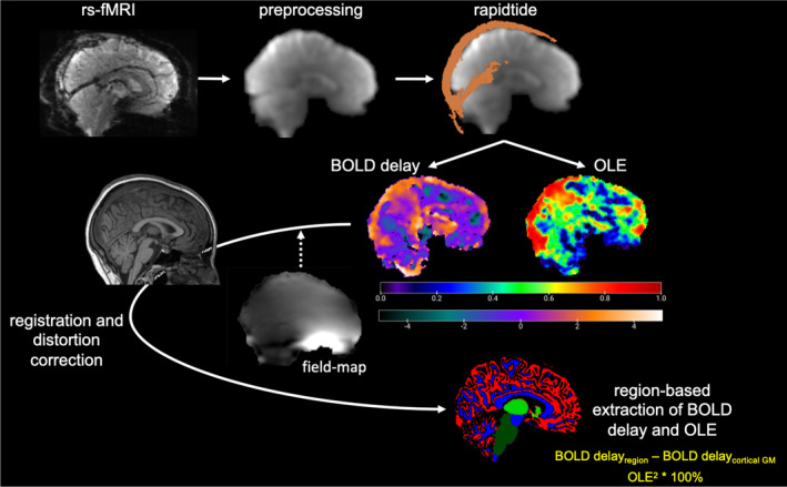
Using resting-state functional magnetic resonance imaging, we investigated brain perfusion and oxygen level estimates in 47 patients (44.4 ± 11.6 years; F:M = 38:9) and 47 individually matched healthy control participants. Group differences were calculated using two-sample t-tests. Multivariable linear regression was used for associations of each regional perfusion and oxygen level measure with cognition and sleepiness measures. Exploratory hazard ratios were calculated for each brain metric with clinical measures.
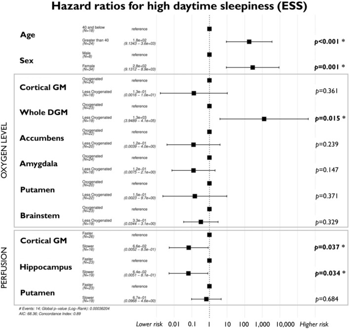
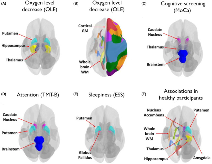
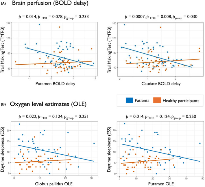
Patients presented with high levels of fatigue (79%) and daytime sleepiness (45%). We found widespread decreased brain oxygen levels, most evident in the white matter (false discovery rate adjusted-p-value (p-) = 0.038) and cortical grey matter (p- = 0.015). Brain perfusion did not differ between patients and healthy participants. However, delayed patient caudate nucleus perfusion was associated with better executive function (p- = 0.008). Delayed perfusion in the cortical grey matter and hippocampus were associated with a reduced risk of daytime sleepiness (hazard ratio (HR) = 0.07, p = 0.037 and HR = 0.06, p = 0.034). Decreased putamen oxygen levels were associated with a reduced risk of poor cognitive outcome (HR = 0.22, p = 0.019). Meanwhile, lower thalamic oxygen levels were associated with a higher risk of cognitive fatigue (HR = 6.29, p = 0.017).
Our findings of lower regional brain blood oxygen levels suggest increased cerebral metabolism in PCS, which potentially holds a compensatory function. These hemodynamic changes were related to symptom severity, possibly representing metabolic adaptations.
新冠后持续存在的神经症状影响多达 10%的患者,并表现为疲劳和认知障碍。基于最近的证据,我们评估了大脑血液动力学变化是否与新冠后综合征(PCS)有关。
我们使用静息态功能磁共振成像,研究了 47 名患者(44.4±11.6 岁;F:M=38:9)和 47 名个体匹配的健康对照参与者的脑灌注和氧水平估计值。使用两样本 t 检验计算组间差异。多元线性回归用于关联每个区域的灌注和氧水平测量与认知和嗜睡测量值。使用临床测量值计算每个脑指标的探索性风险比。
患者表现出高疲劳水平(79%)和白天嗜睡(45%)。我们发现大脑氧水平普遍降低,最明显的是在白质(假发现率调整 p 值(p-)=0.038)和皮质灰质(p-=0.015)。患者和健康参与者之间的脑灌注没有差异。然而,患者尾状核的灌注延迟与更好的执行功能相关(p-=0.008)。皮质灰质和海马的灌注延迟与白天嗜睡风险降低相关(风险比(HR)=0.07,p=0.037 和 HR=0.06,p=0.034)。壳核氧水平降低与认知结局不佳的风险降低相关(HR=0.22,p=0.019)。而丘脑氧水平降低与认知疲劳的风险增加相关(HR=6.29,p=0.017)。
我们发现区域性脑血氧水平降低,提示 PCS 中大脑代谢增加,这可能具有代偿功能。这些血液动力学变化与症状严重程度有关,可能代表代谢适应。