• 文献检索
  • 文档翻译
  • 深度研究
  • 学术资讯
  • Suppr Zotero 插件Zotero 插件
  • 邀请有礼
  • 套餐&价格
  • 历史记录
应用&插件
Suppr Zotero 插件Zotero 插件浏览器插件Mac 客户端Windows 客户端微信小程序
定价
高级版会员购买积分包购买API积分包
服务
文献检索文档翻译深度研究API 文档MCP 服务
关于我们
关于 Suppr公司介绍联系我们用户协议隐私条款
关注我们

Suppr 超能文献

核心技术专利:CN118964589B侵权必究
粤ICP备2023148730 号-1Suppr @ 2026

文献检索

告别复杂PubMed语法,用中文像聊天一样搜索,搜遍4000万医学文献。AI智能推荐,让科研检索更轻松。

立即免费搜索

文件翻译

保留排版,准确专业,支持PDF/Word/PPT等文件格式,支持 12+语言互译。

免费翻译文档

深度研究

AI帮你快速写综述,25分钟生成高质量综述,智能提取关键信息,辅助科研写作。

立即免费体验

高血压可能导致临床前期糖尿病视网膜病变中小动脉壁增厚,而糖尿病则导致临床期视网膜病变中的壁增厚。

Hypertension Likely Drives Arteriolar Wall Thickening in Preclinical Diabetic Retinopathy While Diabetes Drives Wall Thickness in Clinical Retinopathy.

机构信息

Department of Ophthalmology, Feinberg School of Medicine, Northwestern University, Chicago, IL, USA.

出版信息

Transl Vis Sci Technol. 2024 Jun 3;13(6):8. doi: 10.1167/tvst.13.6.8.

DOI:10.1167/tvst.13.6.8
PMID:38874974
原文链接:https://pmc.ncbi.nlm.nih.gov/articles/PMC11182368/
Abstract

PURPOSE

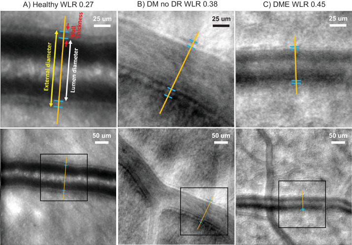
Both hypertension and diabetes are known to increase the wall-to-lumen ratio (WLR) of retinal arterioles, but the differential effects are unknown. Here, we study the timing and relative impact of hypertension versus diabetes on the WLR in diabetic retinopathy (DR) to address this unresolved question.

METHODS

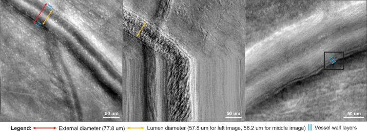
This prospective cross-sectional study compared the retinal arteriolar WLR in 17 healthy eyes, 15 with diabetes but no apparent DR (DM no DR), and 8 with diabetic macular edema (DME) and either nonproliferative or proliferative DR. We imaged each arteriole using adaptive optics scanning laser ophthalmoscopy and measured the WLR using ImageJ. Multiple linear regression (MLR) was performed to estimate the effects of hypertension, diabetes, and age on the WLR.

RESULTS

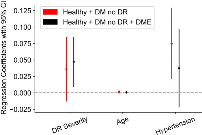
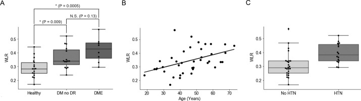
Both subjects with DM no DR and subjects with DME had significantly higher WLR than healthy subjects (0.36 ± 0.08 and 0.42 ± 0.08 vs. 0.29 ± 0.07, 1-way ANOVA P = 0.0009). MLR in healthy subjects and subjects with DM no DR showed hypertension had the strongest effect (regression coefficient = 0.08, P = 0.009), whereas age and diabetes were not significantly correlated with WLR. MLR in all three groups together (healthy, DM no DR, and DME) showed diabetes had the strongest effect (regression coefficient = 0.05, P = 0.02), whereas age and hypertension were not significantly correlated with WLR.

CONCLUSIONS

Hypertension may be an early driver of retinal arteriolar wall thickening in preclinical DR, independent of age or diabetes, whereas changes specific to DR may drive wall thickening in DME and later DR stages.

TRANSLATIONAL RELEVANCE

We offer a framework for understanding the relative contributions of hypertension and diabetes on the vascular wall, and emphasize the importance of hypertension control early in diabetes even before DR onset.

摘要

目的

高血压和糖尿病已知会增加视网膜小动脉的壁腔比(WLR),但尚不清楚其差异影响。在这里,我们研究了高血压与糖尿病对糖尿病视网膜病变(DR)中小动脉壁腔比的时间和相对影响,以解决这个悬而未决的问题。

方法

这项前瞻性横断面研究比较了 17 只健康眼、15 只无明显 DR 的糖尿病眼(DM 无 DR)和 8 只伴有糖尿病性黄斑水肿(DME)且有或无非增生性或增生性 DR 的糖尿病眼的视网膜小动脉 WLR。我们使用自适应光学扫描激光检眼镜对每个小动脉进行成像,并使用 ImageJ 测量 WLR。进行多元线性回归(MLR)以估计高血压、糖尿病和年龄对 WLR 的影响。

结果

DM 无 DR 患者和 DME 患者的 WLR 均明显高于健康受试者(0.36 ± 0.08 和 0.42 ± 0.08 比 0.29 ± 0.07,单因素方差分析 P = 0.0009)。健康受试者和 DM 无 DR 患者的 MLR 显示高血压的影响最强(回归系数 0.08,P = 0.009),而年龄和糖尿病与 WLR 无显著相关性。三组(健康、DM 无 DR 和 DME)的 MLR 均显示糖尿病的影响最强(回归系数 0.05,P = 0.02),而年龄和高血压与 WLR 无显著相关性。

结论

高血压可能是临床前 DR 中小动脉壁增厚的早期驱动因素,独立于年龄或糖尿病,而 DR 特有的变化可能会导致 DME 和更晚期 DR 阶段的壁增厚。

翻译

我们提供了一个理解高血压和糖尿病对血管壁相对影响的框架,并强调了即使在 DR 发生之前,糖尿病早期控制高血压的重要性。

https://cdn.ncbi.nlm.nih.gov/pmc/blobs/3596/11182368/93bea86afc9d/tvst-13-6-8-f004.jpg
https://cdn.ncbi.nlm.nih.gov/pmc/blobs/3596/11182368/36d72dbc1fc5/tvst-13-6-8-f001.jpg
https://cdn.ncbi.nlm.nih.gov/pmc/blobs/3596/11182368/7b41d0f48090/tvst-13-6-8-f002.jpg
https://cdn.ncbi.nlm.nih.gov/pmc/blobs/3596/11182368/8532d157aed9/tvst-13-6-8-f003.jpg
https://cdn.ncbi.nlm.nih.gov/pmc/blobs/3596/11182368/93bea86afc9d/tvst-13-6-8-f004.jpg

相似文献

1
Hypertension Likely Drives Arteriolar Wall Thickening in Preclinical Diabetic Retinopathy While Diabetes Drives Wall Thickness in Clinical Retinopathy.高血压可能导致临床前期糖尿病视网膜病变中小动脉壁增厚,而糖尿病则导致临床期视网膜病变中的壁增厚。
Transl Vis Sci Technol. 2024 Jun 3;13(6):8. doi: 10.1167/tvst.13.6.8.
2
Retinal Vascular Diseases Highlighted by Adaptive Optics Ophthalmoscopy.自适应光学检眼镜凸显的视网膜血管疾病
Rom J Ophthalmol. 2025 Apr-Jun;69(2):235-242. doi: 10.22336/rjo.2025.37.
3
Optical coherence tomography (OCT) for detection of macular oedema in patients with diabetic retinopathy.光学相干断层扫描(OCT)用于检测糖尿病视网膜病变患者的黄斑水肿。
Cochrane Database Syst Rev. 2011 Jul 6(7):CD008081. doi: 10.1002/14651858.CD008081.pub2.
4
Optical coherence tomography (OCT) for detection of macular oedema in patients with diabetic retinopathy.光学相干断层扫描(OCT)用于检测糖尿病视网膜病变患者的黄斑水肿。
Cochrane Database Syst Rev. 2015 Jan 7;1(1):CD008081. doi: 10.1002/14651858.CD008081.pub3.
5
Fenofibrate for diabetic retinopathy.非诺贝特治疗糖尿病性视网膜病变。
Cochrane Database Syst Rev. 2023 Jun 13;6(6):CD013318. doi: 10.1002/14651858.CD013318.pub2.
6
Evaluation of Choroidal Thickness in Type 2 Diabetes Using Spectral-Domain Optical Coherence Tomography.使用频域光学相干断层扫描技术评估2型糖尿病患者的脉络膜厚度
Rom J Ophthalmol. 2025 Apr-Jun;69(2):169-174. doi: 10.22336/rjo.2025.28.
7
Anti-vascular endothelial growth factor for diabetic macular oedema: a network meta-analysis.抗血管内皮生长因子治疗糖尿病性黄斑水肿:一项网状Meta分析。
Cochrane Database Syst Rev. 2017 Jun 22;6(6):CD007419. doi: 10.1002/14651858.CD007419.pub5.
8
Systematic review on urine albumin testing for early detection of diabetic complications.关于尿白蛋白检测用于早期发现糖尿病并发症的系统评价。
Health Technol Assess. 2005 Aug;9(30):iii-vi, xiii-163. doi: 10.3310/hta9300.
9
Anti-VEGF drugs compared with laser photocoagulation for the treatment of diabetic retinopathy: a systematic review and economic analysis.抗血管内皮生长因子药物与激光光凝术治疗糖尿病视网膜病变的比较:系统评价与经济分析
Health Technol Assess. 2025 May 7:1-16. doi: 10.3310/KRWP1264.
10
Anti-vascular endothelial growth factor for diabetic macular oedema: a network meta-analysis.抗血管内皮生长因子治疗糖尿病性黄斑水肿:一项网状Meta分析。
Cochrane Database Syst Rev. 2023 Jun 27;2023(6):CD007419. doi: 10.1002/14651858.CD007419.pub7.

引用本文的文献

1
Advances in materials science for ocular diseases induced by cardiovascular risk factors.心血管危险因素所致眼部疾病的材料科学进展
Front Bioeng Biotechnol. 2025 Jun 27;13:1618232. doi: 10.3389/fbioe.2025.1618232. eCollection 2025.
2
Volumetric Measures of Capillary Nonperfusion on Optical Coherence Tomography Angiography Detect Early Ischemia in Diabetes Without Retinopathy.光学相干断层扫描血管造影术对毛细血管无灌注的容积测量可检测无视网膜病变糖尿病患者的早期缺血。
Invest Ophthalmol Vis Sci. 2025 Apr 1;66(4):2. doi: 10.1167/iovs.66.4.2.

本文引用的文献

1
Imaging the Retinal Vascular Mural Cells In Vivo: Elucidating the Timeline of Their Loss in Diabetic Retinopathy.体内视网膜血管壁细胞成像:阐明糖尿病视网膜病变中其丢失的时间线。
Arterioscler Thromb Vasc Biol. 2024 Feb;44(2):465-476. doi: 10.1161/ATVBAHA.123.320169. Epub 2023 Dec 28.
2
Retinal Arteriolar Wall Remodeling in Diabetes Captured With AOSLO.AOSLO 捕捉到的糖尿病视网膜小动脉壁重构。
Transl Vis Sci Technol. 2023 Nov 1;12(11):16. doi: 10.1167/tvst.12.11.16.
3
Blood pressure control for diabetic retinopathy.糖尿病视网膜病变的血压控制。
Cochrane Database Syst Rev. 2023 Mar 28;3(3):CD006127. doi: 10.1002/14651858.CD006127.pub3.
4
Impact of type 1 diabetes and its duration on wall-to-lumen ratio and blood flow in retinal arterioles.1型糖尿病及其病程对视网膜小动脉壁腔比和血流的影响。
Microvasc Res. 2023 May;147:104499. doi: 10.1016/j.mvr.2023.104499. Epub 2023 Feb 6.
5
Evaluation of Morphological Changes in Retinal Vessels in Type 1 Diabetes Mellitus Patients with the Use of Adaptive Optics.利用自适应光学技术评估1型糖尿病患者视网膜血管的形态学变化
Biomedicines. 2022 Aug 9;10(8):1926. doi: 10.3390/biomedicines10081926.
6
Adaptive optics ophthalmoscopy: a systematic review of vascular biomarkers.自适应光学眼底镜:血管生物标志物的系统评价。
Surv Ophthalmol. 2022 Mar-Apr;67(2):369-387. doi: 10.1016/j.survophthal.2021.05.012. Epub 2021 Jun 6.
7
Association of changes of retinal vessels diameter with ocular blood flow in eyes with diabetic retinopathy.糖尿病视网膜病变患者视网膜血管直径变化与眼血流的关系。
Sci Rep. 2021 Feb 25;11(1):4653. doi: 10.1038/s41598-021-84067-2.
8
RETINAL MICROCIRCULATION INVESTIGATION IN TYPE I AND II DIABETIC PATIENTS WITHOUT RETINOPATHY USING AN ADAPTIVE OPTICS RETINAL CAMERA.使用自适应光学视网膜相机对无视网膜病变的I型和II型糖尿病患者的视网膜微循环进行研究。
Acta Endocrinol (Buchar). 2019 Oct-Dec;15(4):417-422. doi: 10.4183/aeb.2019.417.
9
Early vascular parameters in the micro- and macrocirculation in type 2 diabetes.2 型糖尿病患者的微血管和大血管中的早期血管参数。
Cardiovasc Diabetol. 2018 Sep 19;17(1):128. doi: 10.1186/s12933-018-0770-4.
10
Comparison between invasive and noninvasive techniques of evaluation of microvascular structural alterations.比较评估微血管结构改变的侵袭性和非侵袭性技术。
J Hypertens. 2018 May;36(5):1154-1163. doi: 10.1097/HJH.0000000000001677.