MSIII Joan C. Edwards School of Medicine, Huntington, WV, USA.
Department of Dermatology, University of Minnesota, Minneapolis, MN, USA.
Drugs R D. 2024 Jun;24(2):353-357. doi: 10.1007/s40268-024-00461-x. Epub 2024 Jun 15.
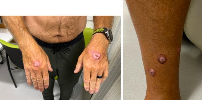
PD-1 inhibitors have revolutionized cancer therapies and are being used to treat an expanding array of cancers. To best serve patients, clinicians should be familiar with the spectrum of skin manifestations associated with PD-1 inhibitor therapy. Here, we report a unique case of hypertrophic lichen planus (HLP) in a 64-year-old man treated with pembrolizumab; the presentation initially suggested a squamous cell carcinoma (SCC) morphology, then evolved into a morphology more typical of hypertrophic lichen planus. This case underscores the need for caution in diagnosing eruptive SCCs associated with PD-1 inhibitor therapy. In such instances, maintaining a high suspicion for lichenoid reactions as sequelae of PD-1 inhibitor treatment and starting an empiric trial of therapy for lichenoid dermatitis may be warranted to ensure timely management of lesions.
We describe a case of hypertrophic lichen planus mimicking squamous cell carcinoma in the setting of PD-1 inhibitory therapy with pembrolizumab. A PubMed literature review was conducted to identify other cases and determine the incidence of lichenoid reactions imitating squamous cell carcinoma in the setting of PD-1 inhibitor use.
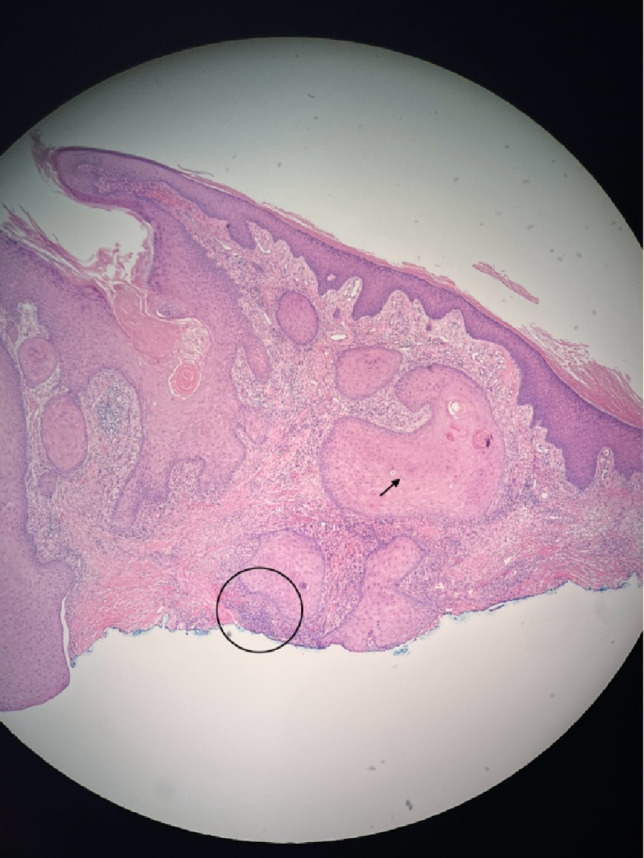
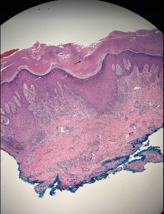
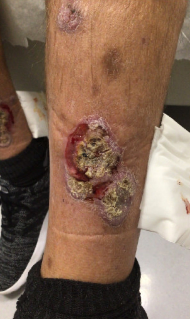
Our case is one of the few available pieces of literature describing eruptive hypertrophic lichen planus imitating SCC in the setting of PD-1 inhibitor use. Initial skin nodule biopsy appeared histologically compatible with squamous cell carcinoma. Repeat biopsy of the skin lesions revealed histological features consistent with hypertrophic lichen planus. Over time, lower extremity lesions evolved into a more typical appearance of hypertrophic lichen planus. Treatment with topical 0.05% clobetasol ointment and oral acitretin 25 mg led to complete resolution of lesions within 2-3 months.
This case underscores the significance of maintaining vigilance for lichenoid reactions as potential sequelae of PD-1 inhibitor therapy. It highlights the variability in initial presentation and the potential for lesions to transform over time. Timely recognition and appropriate management, including high-potency topical corticosteroids and oral acitretin, are crucial for achieving favorable outcomes in patients experiencing such reactions. More studies are necessary to fully analyze the rate of HLP occurrence as a consequence of PD-1 inhibitor use.
PD-1 抑制剂彻底革新了癌症治疗方法,并被广泛应用于多种癌症的治疗。为了更好地服务患者,临床医生应熟悉 PD-1 抑制剂治疗相关的皮肤表现谱。在这里,我们报告了一例使用派姆单抗治疗的 64 岁男性发生肥厚性扁平苔藓(HLP)的独特病例;该患者的最初表现提示为鳞状细胞癌(SCC)形态,随后演变为更典型的肥厚性扁平苔藓形态。该病例强调了在诊断与 PD-1 抑制剂治疗相关的爆发性 SCC 时需保持谨慎。在这种情况下,维持对 PD-1 抑制剂治疗后继发的苔藓样反应的高度怀疑,并开始进行经验性的苔藓样皮炎治疗试验,可能有助于及时管理病变。
我们描述了一例在使用派姆单抗进行 PD-1 抑制治疗的情况下,模仿鳞状细胞癌的肥厚性扁平苔藓病例。我们进行了一项 PubMed 文献回顾,以确定其他病例,并确定 PD-1 抑制剂使用情况下模仿鳞状细胞癌的苔藓样反应的发生率。
我们的病例是少数描述在 PD-1 抑制剂使用情况下爆发性肥厚性扁平苔藓模仿 SCC 的文献之一。初始皮肤结节活检在组织学上与鳞状细胞癌相符。皮肤病变的重复活检显示出与肥厚性扁平苔藓一致的组织学特征。随着时间的推移,下肢病变演变为更典型的肥厚性扁平苔藓外观。局部使用 0.05%卤倍他索软膏和口服阿维 A 25mg 治疗导致病变在 2-3 个月内完全消退。
该病例强调了对 PD-1 抑制剂治疗潜在的苔藓样反应保持警惕的重要性。它突出了初始表现的可变性和病变随时间推移而变化的可能性。及时识别和适当管理,包括高效局部皮质类固醇和口服阿维 A,对于患有此类反应的患者获得良好的治疗结果至关重要。需要更多的研究来全面分析 PD-1 抑制剂使用导致 HLP 发生的发生率。