• 文献检索
  • 文档翻译
  • 深度研究
  • 学术资讯
  • Suppr Zotero 插件Zotero 插件
  • 邀请有礼
  • 套餐&价格
  • 历史记录
应用&插件
Suppr Zotero 插件Zotero 插件浏览器插件Mac 客户端Windows 客户端微信小程序
定价
高级版会员购买积分包购买API积分包
服务
文献检索文档翻译深度研究API 文档MCP 服务
关于我们
关于 Suppr公司介绍联系我们用户协议隐私条款
关注我们

Suppr 超能文献

核心技术专利:CN118964589B侵权必究
粤ICP备2023148730 号-1Suppr @ 2026

文献检索

告别复杂PubMed语法,用中文像聊天一样搜索,搜遍4000万医学文献。AI智能推荐,让科研检索更轻松。

立即免费搜索

文件翻译

保留排版,准确专业,支持PDF/Word/PPT等文件格式,支持 12+语言互译。

免费翻译文档

深度研究

AI帮你快速写综述,25分钟生成高质量综述,智能提取关键信息,辅助科研写作。

立即免费体验

利用 TRACERx-PHLEX 进行深度细胞表型分析和多重成像的空间分析。

Deep cell phenotyping and spatial analysis of multiplexed imaging with TRACERx-PHLEX.

机构信息

Cancer Evolution and Genome Instability Laboratory, The Francis Crick Institute, London, UK.

Cancer Research UK Lung Cancer Centre of Excellence, University College London Cancer Institute, London, UK.

出版信息

Nat Commun. 2024 Jun 15;15(1):5135. doi: 10.1038/s41467-024-48870-5.

DOI:10.1038/s41467-024-48870-5
PMID:38879602
原文链接:https://pmc.ncbi.nlm.nih.gov/articles/PMC11180132/
Abstract

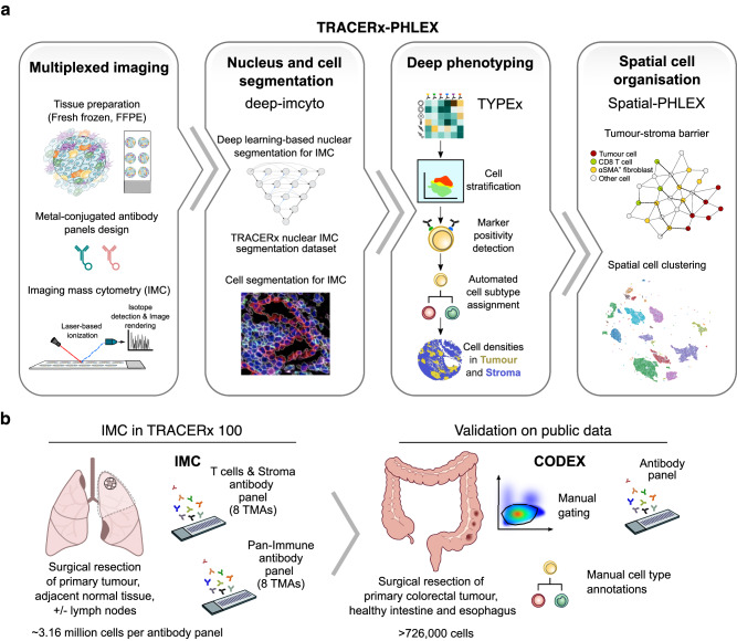
The growing scale and dimensionality of multiplexed imaging require reproducible and comprehensive yet user-friendly computational pipelines. TRACERx-PHLEX performs deep learning-based cell segmentation (deep-imcyto), automated cell-type annotation (TYPEx) and interpretable spatial analysis (Spatial-PHLEX) as three independent but interoperable modules. PHLEX generates single-cell identities, cell densities within tissue compartments, marker positivity calls and spatial metrics such as cellular barrier scores, along with summary graphs and spatial visualisations. PHLEX was developed using imaging mass cytometry (IMC) in the TRACERx study, validated using published Co-detection by indexing (CODEX), IMC and orthogonal data and benchmarked against state-of-the-art approaches. We evaluated its use on different tissue types, tissue fixation conditions, image sizes and antibody panels. As PHLEX is an automated and containerised Nextflow pipeline, manual assessment, programming skills or pathology expertise are not essential. PHLEX offers an end-to-end solution in a growing field of highly multiplexed data and provides clinically relevant insights.

摘要

多指标成像的规模和维度不断扩大,这就需要可重复、全面且用户友好的计算流程。TRACERx-PHLEX 作为三个独立但可互操作的模块,执行基于深度学习的细胞分割(deep-imcyto)、自动细胞类型注释(TYPEx)和可解释的空间分析(Spatial-PHLEX)。PHLEX 生成单细胞身份、组织隔室中细胞密度、标记物阳性调用以及细胞屏障评分等空间指标,同时生成摘要图和空间可视化。PHLEX 是在 TRACERx 研究中使用成像质谱细胞术(IMC)开发的,使用已发表的 Co-detection by indexing (CODEX)、IMC 和正交数据进行了验证,并与最先进的方法进行了基准测试。我们评估了它在不同组织类型、组织固定条件、图像大小和抗体面板上的使用情况。由于 PHLEX 是一个自动化和容器化的 Nextflow 流程,因此不需要手动评估、编程技能或病理学专业知识。PHLEX 在高度多指标数据不断增长的领域提供了端到端的解决方案,并提供了临床相关的见解。

https://cdn.ncbi.nlm.nih.gov/pmc/blobs/014e/11180132/a2654d2dac78/41467_2024_48870_Fig7_HTML.jpg
https://cdn.ncbi.nlm.nih.gov/pmc/blobs/014e/11180132/ada49c59b621/41467_2024_48870_Fig1_HTML.jpg
https://cdn.ncbi.nlm.nih.gov/pmc/blobs/014e/11180132/cd11c228dc50/41467_2024_48870_Fig2_HTML.jpg
https://cdn.ncbi.nlm.nih.gov/pmc/blobs/014e/11180132/62782bb70fe2/41467_2024_48870_Fig3_HTML.jpg
https://cdn.ncbi.nlm.nih.gov/pmc/blobs/014e/11180132/0704fe6c2a05/41467_2024_48870_Fig4_HTML.jpg
https://cdn.ncbi.nlm.nih.gov/pmc/blobs/014e/11180132/29ad65c95bee/41467_2024_48870_Fig5_HTML.jpg
https://cdn.ncbi.nlm.nih.gov/pmc/blobs/014e/11180132/5fc1258f601e/41467_2024_48870_Fig6_HTML.jpg
https://cdn.ncbi.nlm.nih.gov/pmc/blobs/014e/11180132/a2654d2dac78/41467_2024_48870_Fig7_HTML.jpg

相似文献

1
Deep cell phenotyping and spatial analysis of multiplexed imaging with TRACERx-PHLEX.利用 TRACERx-PHLEX 进行深度细胞表型分析和多重成像的空间分析。
Nat Commun. 2024 Jun 15;15(1):5135. doi: 10.1038/s41467-024-48870-5.
2
CellSeg: a robust, pre-trained nucleus segmentation and pixel quantification software for highly multiplexed fluorescence images.CellSeg:一款用于高度多重荧光图像的强大的、预训练的细胞核分割和像素定量软件。
BMC Bioinformatics. 2022 Jan 18;23(1):46. doi: 10.1186/s12859-022-04570-9.
3
An end-to-end workflow for multiplexed image processing and analysis.用于多重图像处理和分析的端到端工作流程。
Nat Protoc. 2023 Nov;18(11):3565-3613. doi: 10.1038/s41596-023-00881-0. Epub 2023 Oct 10.
4
Accurate single-molecule spot detection for image-based spatial transcriptomics with weakly supervised deep learning.基于弱监督深度学习的图像空间转录组学中单分子斑点的精确检测。
Cell Syst. 2024 May 15;15(5):475-482.e6. doi: 10.1016/j.cels.2024.04.006.
5
TITAN: An end-to-end data analysis environment for the Hyperion™ imaging system.TITAN:适用于Hyperion™成像系统的端到端数据分析环境。
Cytometry A. 2022 May;101(5):423-433. doi: 10.1002/cyto.a.24535. Epub 2022 Jan 28.
6
A review on deep learning applications in highly multiplexed tissue imaging data analysis.深度学习在高度多重组织成像数据分析中的应用综述。
Front Bioinform. 2023 Jul 26;3:1159381. doi: 10.3389/fbinf.2023.1159381. eCollection 2023.
7
Cytokit: a single-cell analysis toolkit for high dimensional fluorescent microscopy imaging.细胞分析工具包:用于高维荧光显微镜成像的单细胞分析工具包。
BMC Bioinformatics. 2019 Sep 2;20(1):448. doi: 10.1186/s12859-019-3055-3.
8
Tribus: semi-automated discovery of cell identities and phenotypes from multiplexed imaging and proteomic data.Tribus:从多重成像和蛋白质组学数据中半自动发现细胞身份和表型。
Bioinformatics. 2025 Mar 4;41(3). doi: 10.1093/bioinformatics/btaf082.
9
An open-source solution for advanced imaging flow cytometry data analysis using machine learning.一种使用机器学习进行高级成像流式细胞术数据分析的开源解决方案。
Methods. 2017 Jan 1;112:201-210. doi: 10.1016/j.ymeth.2016.08.018. Epub 2016 Sep 2.
10
A novel process for H&E, immunofluorescence, and imaging mass cytometry on a single slide with a concise analytics pipeline.一种在单张载玻片上进行苏木精和伊红染色、免疫荧光及成像质谱流式细胞术的新型方法,且具备简洁的分析流程。
Cytometry A. 2023 Dec;103(12):1010-1018. doi: 10.1002/cyto.a.24789. Epub 2023 Sep 19.

引用本文的文献

1
Deconvolution of cell types and states in spatial multiomics utilizing TACIT.利用TACIT对空间多组学中的细胞类型和状态进行反卷积分析。
Nat Commun. 2025 Apr 21;16(1):3747. doi: 10.1038/s41467-025-58874-4.
2
Large-volume fully automated cell reconstruction generates a cell atlas of plant tissues.大体积全自动化细胞重建生成了植物组织的细胞图谱。
Plant Cell. 2024 Sep 16;36(12):4840-61. doi: 10.1093/plcell/koae250.

本文引用的文献

1
Spatial Architecture of Myeloid and T Cells Orchestrates Immune Evasion and Clinical Outcome in Lung Cancer.髓系和 T 细胞的空间结构调控肺癌的免疫逃逸和临床结局。
Cancer Discov. 2024 Jun 3;14(6):1018-1047. doi: 10.1158/2159-8290.CD-23-1380.
2
CellCharter reveals spatial cell niches associated with tissue remodeling and cell plasticity.CellCharter 揭示了与组织重塑和细胞可塑性相关的空间细胞龛。
Nat Genet. 2024 Jan;56(1):74-84. doi: 10.1038/s41588-023-01588-4. Epub 2023 Dec 8.
3
An end-to-end workflow for multiplexed image processing and analysis.
用于多重图像处理和分析的端到端工作流程。
Nat Protoc. 2023 Nov;18(11):3565-3613. doi: 10.1038/s41596-023-00881-0. Epub 2023 Oct 10.
4
Unsupervised discovery of tissue architecture in multiplexed imaging.无监督发现多重成像中的组织架构。
Nat Methods. 2022 Dec;19(12):1653-1661. doi: 10.1038/s41592-022-01657-2. Epub 2022 Oct 31.
5
Annotation of spatially resolved single-cell data with STELLAR.利用 STELLAR 对空间分辨单细胞数据进行注释。
Nat Methods. 2022 Nov;19(11):1411-1418. doi: 10.1038/s41592-022-01651-8. Epub 2022 Oct 24.
6
Scalable and model-free detection of spatial patterns and colocalization.可扩展且无模型的空间模式和共定位检测。
Genome Res. 2022 Sep;32(9):1736-1745. doi: 10.1101/gr.276851.122. Epub 2022 Sep 9.
7
Single-cell analyses define a continuum of cell state and composition changes in the malignant transformation of polyps to colorectal cancer.单细胞分析定义了息肉恶变为结直肠癌过程中细胞状态和组成变化的连续体。
Nat Genet. 2022 Jul;54(7):985-995. doi: 10.1038/s41588-022-01088-x. Epub 2022 Jun 20.
8
Identification of cell types in multiplexed in situ images by combining protein expression and spatial information using CELESTA.通过结合蛋白表达和空间信息使用 CELESTA 对多重原位图像中的细胞类型进行鉴定。
Nat Methods. 2022 Jun;19(6):759-769. doi: 10.1038/s41592-022-01498-z. Epub 2022 Jun 2.
9
Multiplexed imaging mass cytometry of the chemokine milieus in melanoma characterizes features of the response to immunotherapy.免疫治疗应答特征的黑色素瘤趋化因子微环境的多重成像质谱细胞术分析。
Sci Immunol. 2022 Apr;7(70):eabk1692. doi: 10.1126/sciimmunol.abk1692. Epub 2022 Apr 1.
10
A SIMPLI (Single-cell Identification from MultiPLexed Images) approach for spatially-resolved tissue phenotyping at single-cell resolution.一种基于 SIMPLI(从多重成像中鉴定单细胞)的方法,可实现单细胞分辨率的空间分辨组织表型分析。
Nat Commun. 2022 Feb 9;13(1):781. doi: 10.1038/s41467-022-28470-x.