Lund Leah M, Marchi Angelina N, Alderfer Laura, Hall Eva, Hammer Jacob, Trull Keelan J, Hanjaya-Putra Donny, White Katharine A
Department of Chemistry and Biochemistry, University of Notre Dame, 251 Nieuwland Science Hall, Notre Dame, IN 46556 USA.
Harper Cancer Research Institute, University of Notre Dame, 1234 N. Notre Dame Avenue, South Bend, IN 46617 USA.
bioRxiv. 2024 Jun 4:2024.06.04.597454. doi: 10.1101/2024.06.04.597454.
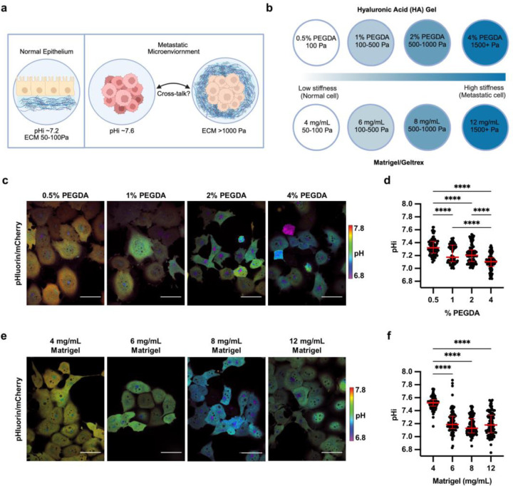
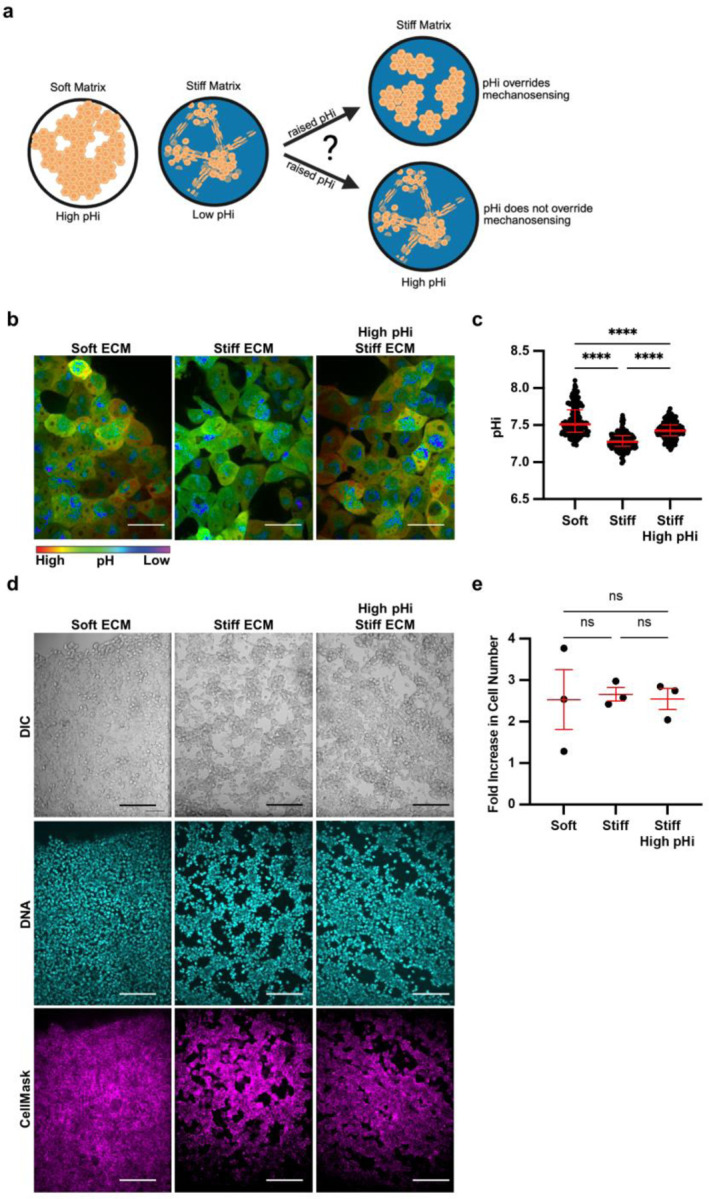
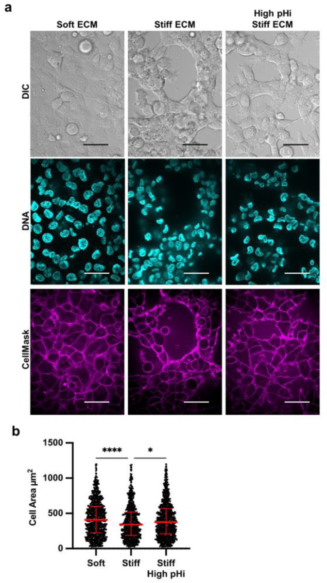
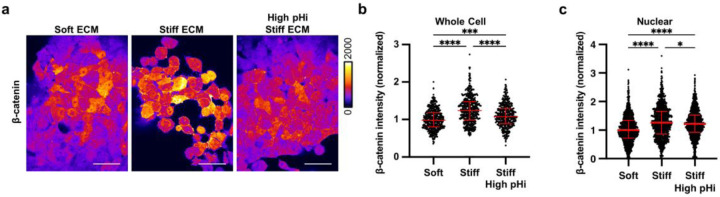
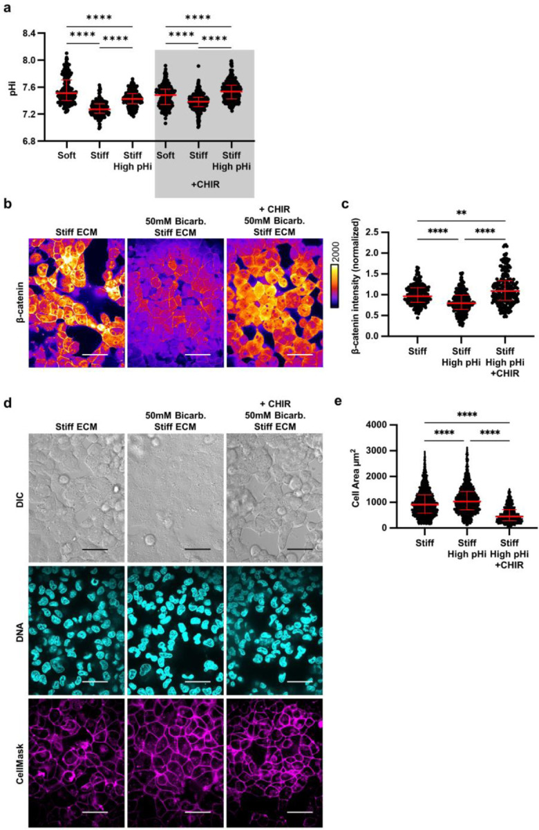
Dysregulated intracellular pH (pHi) dynamics and an altered tumor microenvironment have emerged as drivers of cancer cell phenotypes. However, the molecular integration between the physical properties of the microenvironment and dynamic intracellular signaling responses remains unclear. Here, we use two metastatic cell models, one breast and one lung, to assess pHi response to varying extracellular matrix (ECM) stiffness. To experimentally model ECM stiffening, we use two tunable-stiffness hydrogel systems: Matrigel and hyaluronic acid (HA) gels, which mimic the increased protein secretion and crosslinking associated with ECM stiffening. We find that single-cell pHi decreases with increased ECM stiffness in both hydrogel systems and both metastatic cell types. We also observed that stiff ECM promotes vasculogenic mimicry (VM), a phenotype associated with metastasis and resistance. Importantly, we show that decreased pHi is both a necessary and sufficient mediator of VM, as raising pHi on stiff ECM reduces VM phenotypes and lowering pHi on soft ECM drives VM. We characterize β-catenin as a pH-dependent molecular mediator of pH-dependent VM, where stiffness-driven changes in β-catenin abundance can be overridden by increased pHi. We uncover a dynamic relationship between matrix stiffness and pHi, thus suggesting pHi dynamics can override mechanosensitive cell responses to the extracellular microenvironment.
细胞内pH值(pHi)动态失调以及肿瘤微环境改变已成为癌细胞表型的驱动因素。然而,微环境物理特性与细胞内动态信号反应之间的分子整合仍不清楚。在此,我们使用两种转移细胞模型,一种是乳腺癌细胞模型,另一种是肺癌细胞模型,来评估pHi对不同细胞外基质(ECM)硬度的反应。为了通过实验模拟ECM硬化,我们使用两种可调硬度水凝胶系统:基质胶和透明质酸(HA)凝胶,它们模拟了与ECM硬化相关的蛋白质分泌增加和交联。我们发现,在两种水凝胶系统和两种转移细胞类型中,单细胞pHi均随着ECM硬度增加而降低。我们还观察到,坚硬的ECM促进血管生成拟态(VM),这是一种与转移和耐药相关的表型。重要的是,我们表明pHi降低是VM的必要且充分介导因素,因为在坚硬ECM上提高pHi可减少VM表型,而在柔软ECM上降低pHi则会驱动VM。我们将β-连环蛋白表征为pH依赖性VM的pH依赖性分子介导物,其中硬度驱动的β-连环蛋白丰度变化可被升高的pHi所抵消。我们揭示了基质硬度与pHi之间的动态关系,从而表明pHi动态变化可超越细胞对细胞外微环境的机械敏感反应。