Szachowicz Peter J, Wohlford-Lenane Christine, Heinen Cobey J, Ghimire Shreya, Xue Biyun, Boly Timothy J, Verma Abhishek, MašinoviĆ Leila, Bermick Jennifer R, Perlman Stanley, Meyerholz David K, Pezzulo Alejandro A, Zhang Yuzhou, Smith Richard J H, McCray Paul B
Department of Internal Medicine, The University of Iowa, Division of Pulmonary, Critical Care, and Occupational Medicine, Iowa City, IA, 52242.
Stead Family Department of Pediatrics, The University of Iowa, Iowa City, IA, 52242.
bioRxiv. 2024 Jun 3:2024.05.31.596892. doi: 10.1101/2024.05.31.596892.
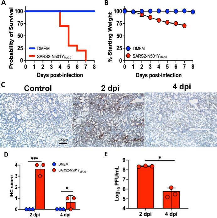
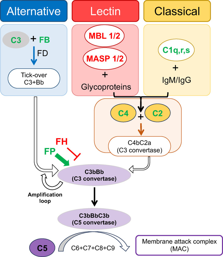
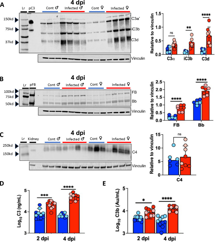
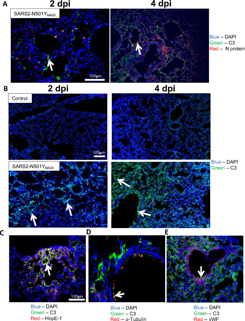
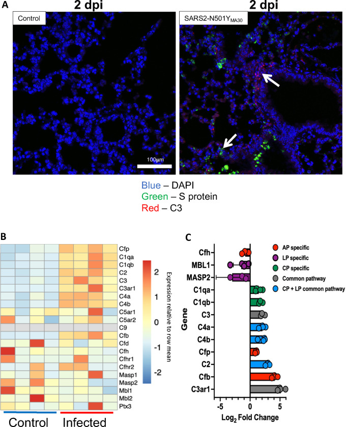
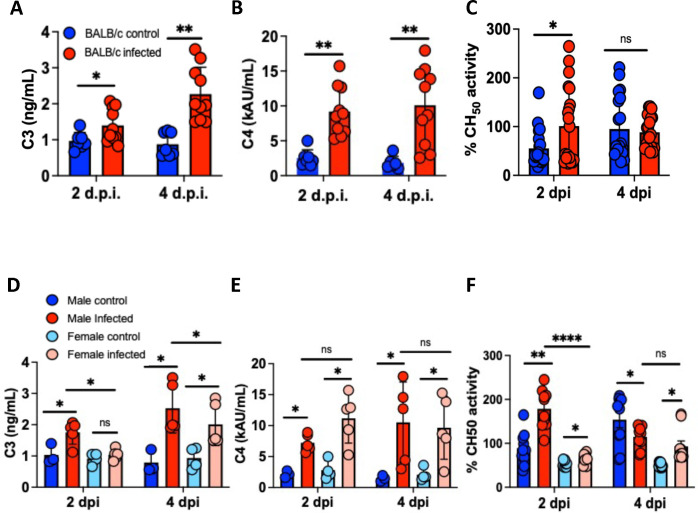
Evidence from in vitro studies and observational human disease data suggest the complement system plays a significant role in SARS-CoV-2 pathogenesis, although how complement dysregulation develops in patients with severe COVID-19 is unknown. Here, using a mouse-adapted SARS-CoV-2 virus (SARS2-N501Y) and a mouse model of severe COVID-19, we identify significant serologic and pulmonary complement activation following infection. We observed C3 activation in airway and alveolar epithelia, and in pulmonary vascular endothelia. Our evidence suggests that while the alternative pathway is the primary route of complement activation, components of both the alternative and classical pathways are produced locally by respiratory epithelial cells following infection, and increased in primary cultures of human airway epithelia in response to cytokine exposure. This locally generated complement response appears to precede and subsequently drive lung injury and inflammation. Results from this mouse model recapitulate findings in humans, which suggest sex-specific variance in complement activation, with predilection for increased C3 activity in males, a finding that may correlate with more severe disease. Our findings indicate that complement activation is a defining feature of severe COVID-19 in mice and lay the foundation for further investigation into the role of complement in COVID-19.
体外研究和人类疾病观察数据的证据表明,补体系统在SARS-CoV-2发病机制中起重要作用,尽管严重COVID-19患者的补体失调是如何发生的尚不清楚。在此,我们使用适应小鼠的SARS-CoV-2病毒(SARS2-N501Y)和严重COVID-19小鼠模型,确定感染后血清学和肺部补体的显著激活。我们观察到气道和肺泡上皮以及肺血管内皮中的C3激活。我们的证据表明,虽然替代途径是补体激活的主要途径,但替代途径和经典途径的成分在感染后均由呼吸道上皮细胞在局部产生,并在人气道上皮原代培养物中因细胞因子暴露而增加。这种局部产生的补体反应似乎先于并随后驱动肺损伤和炎症。该小鼠模型的结果重现了人类的研究结果,提示补体激活存在性别特异性差异,男性的C3活性增加更为明显,这一发现可能与更严重的疾病相关。我们的研究结果表明,补体激活是小鼠严重COVID-19的一个决定性特征,并为进一步研究补体在COVID-19中的作用奠定了基础。