• 文献检索
  • 文档翻译
  • 深度研究
  • 学术资讯
  • Suppr Zotero 插件Zotero 插件
  • 邀请有礼
  • 套餐&价格
  • 历史记录
应用&插件
Suppr Zotero 插件Zotero 插件浏览器插件Mac 客户端Windows 客户端微信小程序
定价
高级版会员购买积分包购买API积分包
服务
文献检索文档翻译深度研究API 文档MCP 服务
关于我们
关于 Suppr公司介绍联系我们用户协议隐私条款
关注我们

Suppr 超能文献

核心技术专利:CN118964589B侵权必究
粤ICP备2023148730 号-1Suppr @ 2026

文献检索

告别复杂PubMed语法,用中文像聊天一样搜索,搜遍4000万医学文献。AI智能推荐,让科研检索更轻松。

立即免费搜索

文件翻译

保留排版,准确专业,支持PDF/Word/PPT等文件格式,支持 12+语言互译。

免费翻译文档

深度研究

AI帮你快速写综述,25分钟生成高质量综述,智能提取关键信息,辅助科研写作。

立即免费体验

创伤性轴索损伤在早期 MRI 中的预后意义:特隆赫姆 TAI-MRI 分级和定量模型。

The prognostic importance of traumatic axonal injury on early MRI: the Trondheim TAI-MRI grading and quantitative models.

机构信息

Department of Circulation and Medical Imaging, Faculty of Medicine and Health Sciences, Norwegian University of Science and Technology (NTNU), 7491, Trondheim, Norway.

Department of Radiology, Vestre Viken Hospital Trust, Drammen Hospital, 3004, Drammen, Norway.

出版信息

Eur Radiol. 2024 Dec;34(12):8015-8029. doi: 10.1007/s00330-024-10841-1. Epub 2024 Jun 19.

DOI:10.1007/s00330-024-10841-1
PMID:38896232
原文链接:https://pmc.ncbi.nlm.nih.gov/articles/PMC11557676/
Abstract

OBJECTIVES

We analysed magnetic resonance imaging (MRI) findings after traumatic brain injury (TBI) aiming to improve the grading of traumatic axonal injury (TAI) to better reflect the outcome.

METHODS

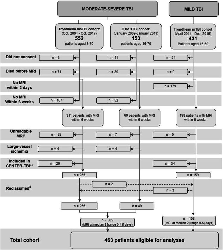
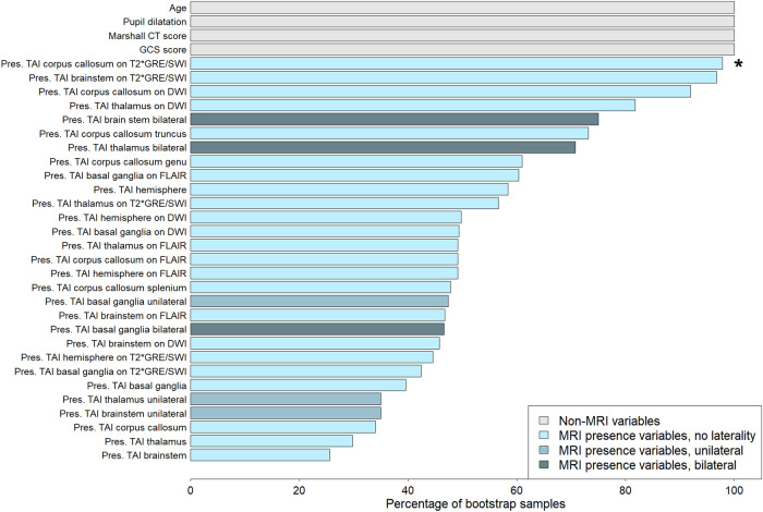
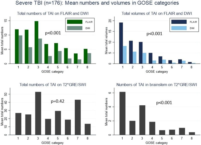
Four-hundred sixty-three patients (8-70 years) with mild (n = 158), moderate (n = 129), or severe (n = 176) TBI and early MRI were prospectively included. TAI presence, numbers, and volumes at predefined locations were registered on fluid-attenuated inversion recovery (FLAIR) and diffusion-weighted imaging, and presence and numbers on T2*GRE/SWI. Presence and volumes of contusions were registered on FLAIR. We assessed the outcome with the Glasgow Outcome Scale Extended. Multivariable logistic and elastic-net regression analyses were performed.

RESULTS

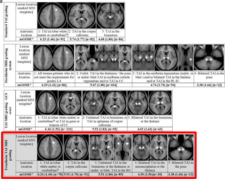
The presence of TAI differed between mild (6%), moderate (70%), and severe TBI (95%). In severe TBI, bilateral TAI in mesencephalon or thalami and bilateral TAI in pons predicted worse outcomes and were defined as the worst grades (4 and 5, respectively) in the Trondheim TAI-MRI grading. The Trondheim TAI-MRI grading performed better than the standard TAI grading in severe TBI (pseudo-R 0.19 vs. 0.16). In moderate-severe TBI, quantitative models including both FLAIR volume of TAI and contusions performed best (pseudo-R 0.19-0.21). In patients with mild TBI or Glasgow Coma Scale (GCS) score 13, models with the volume of contusions performed best (pseudo-R 0.25-0.26).

CONCLUSIONS

We propose the Trondheim TAI-MRI grading (grades 1-5) with bilateral TAI in mesencephalon or thalami, and bilateral TAI in pons as the worst grades. The predictive value was highest for the quantitative models including FLAIR volume of TAI and contusions (GCS score <13) or FLAIR volume of contusions (GCS score ≥ 13), which emphasise artificial intelligence as a potentially important future tool.

CLINICAL RELEVANCE STATEMENT

The Trondheim TAI-MRI grading reflects patient outcomes better in severe TBI than today's standard TAI grading and can be implemented after external validation. The prognostic importance of volumetric models is promising for future use of artificial intelligence technologies.

KEY POINTS

Traumatic axonal injury (TAI) is an important injury type in all TBI severities. Studies demonstrating which MRI findings that can serve as future biomarkers are highly warranted. This study proposes the most optimal MRI models for predicting patient outcome at 6 months after TBI; one updated pragmatic model and a volumetric model. The Trondheim TAI-MRI grading, in severe TBI, reflects patient outcome better than today's standard grading of TAI and the prognostic importance of volumetric models in all severities of TBI is promising for future use of AI.

摘要

目的

我们分析了外伤性脑损伤(TBI)后的磁共振成像(MRI)结果,旨在改善外伤性轴索损伤(TAI)的分级,以更好地反映结果。

方法

前瞻性纳入了 463 例(8-70 岁)轻度(n=158)、中度(n=129)或重度(n=176)TBI 且早期 MRI 患者。在液体衰减反转恢复(FLAIR)和弥散加权成像上记录 TAI 的存在、数量和体积,在 T2*GRE/SWI 上记录 TAI 的存在和数量。在 FLAIR 上记录挫伤的存在和体积。我们使用格拉斯哥预后量表扩展版评估了预后。进行了多变量逻辑和弹性网回归分析。

结果

在轻度(6%)、中度(70%)和重度 TBI(95%)中,TAI 的存在存在差异。在重度 TBI 中,中脑或丘脑的双侧 TAI 和脑桥的双侧 TAI 预测预后更差,分别被定义为 Trondheim TAI-MRI 分级的最差等级(分别为 4 级和 5 级)。在重度 TBI 中,Trondheim TAI-MRI 分级的表现优于标准 TAI 分级(伪 R 0.19 与 0.16)。在中重度 TBI 中,包括 FLAIR 上 TAI 体积和挫伤的定量模型表现最佳(伪 R 0.19-0.21)。在轻度 TBI 或格拉斯哥昏迷量表(GCS)评分 13 的患者中,包含挫伤体积的模型表现最佳(伪 R 0.25-0.26)。

结论

我们提出了 Trondheim TAI-MRI 分级(1-5 级),中脑或丘脑的双侧 TAI 和脑桥的双侧 TAI 为最差等级。包括 FLAIR 上 TAI 体积和挫伤的定量模型或 FLAIR 上挫伤体积(GCS 评分≥13)的预测价值最高,这强调了人工智能作为一种潜在的重要未来工具。

临床相关性声明

与目前的标准 TAI 分级相比,Trondheim TAI-MRI 分级在重度 TBI 中能更好地反映患者预后,且可在外部验证后实施。容积模型的预后重要性为未来使用人工智能技术提供了希望。

关键点

外伤性轴索损伤(TAI)是所有 TBI 严重程度中的一种重要损伤类型。迫切需要研究证明哪些 MRI 发现可以作为未来的生物标志物。本研究提出了预测 TBI 后 6 个月患者预后的最佳 MRI 模型;一个更新的实用模型和一个体积模型。在重度 TBI 中,Trondheim TAI-MRI 分级比目前的 TAI 分级更能反映患者的预后,且所有严重程度的 TBI 中容积模型的预后重要性对未来人工智能的应用具有很大的潜力。

https://cdn.ncbi.nlm.nih.gov/pmc/blobs/c791/11557676/cd6dd35b846a/330_2024_10841_Fig4a_HTML.jpg
https://cdn.ncbi.nlm.nih.gov/pmc/blobs/c791/11557676/a5c84b9fb081/330_2024_10841_Fig1_HTML.jpg
https://cdn.ncbi.nlm.nih.gov/pmc/blobs/c791/11557676/6d3fe4481548/330_2024_10841_Fig2_HTML.jpg
https://cdn.ncbi.nlm.nih.gov/pmc/blobs/c791/11557676/af37ff124a37/330_2024_10841_Fig3_HTML.jpg
https://cdn.ncbi.nlm.nih.gov/pmc/blobs/c791/11557676/cd6dd35b846a/330_2024_10841_Fig4a_HTML.jpg

相似文献

1
The prognostic importance of traumatic axonal injury on early MRI: the Trondheim TAI-MRI grading and quantitative models.创伤性轴索损伤在早期 MRI 中的预后意义:特隆赫姆 TAI-MRI 分级和定量模型。
Eur Radiol. 2024 Dec;34(12):8015-8029. doi: 10.1007/s00330-024-10841-1. Epub 2024 Jun 19.
2
Lesion Frequency Distribution Maps of Traumatic Axonal Injury on Early Magnetic Resonance Imaging After Moderate and Severe Traumatic Brain Injury and Associations to 12 Months Outcome.创伤性轴索损伤病灶频率分布图谱在中重度创伤性脑损伤后早期磁共振成像中的表现及其与 12 个月结局的相关性。
J Neurotrauma. 2024 Aug;41(15-16):1901-1913. doi: 10.1089/neu.2023.0534. Epub 2024 Apr 29.
3
Traumatic axonal injury: the prognostic value of lesion load in corpus callosum, brain stem, and thalamus in different magnetic resonance imaging sequences.创伤性轴索损伤:不同磁共振成像序列中胼胝体、脑干和丘脑病变负荷的预后价值
J Neurotrauma. 2014 Sep 1;31(17):1486-96. doi: 10.1089/neu.2013.3258. Epub 2014 Jul 1.
4
Traumatic axonal injury on clinical MRI: association with the Glasgow Coma Scale score at scene of injury or at admission and prolonged posttraumatic amnesia.临床MRI上的创伤性轴索损伤:与受伤现场或入院时的格拉斯哥昏迷量表评分及创伤后遗忘期延长的相关性
J Neurosurg. 2020 Oct 23;135(2):562-573. doi: 10.3171/2020.6.JNS20112. Print 2021 Aug 1.
5
A longitudinal MRI study of traumatic axonal injury in patients with moderate and severe traumatic brain injury.一项关于中重度颅脑创伤患者创伤性轴索损伤的纵向 MRI 研究。
J Neurol Neurosurg Psychiatry. 2012 Dec;83(12):1193-200. doi: 10.1136/jnnp-2012-302644. Epub 2012 Aug 29.
6
Traumatic axonal injury: is the prognostic information produced by conventional MRI and DTI complementary or supplementary?创伤性轴索损伤:常规 MRI 和 DTI 提供的预后信息是互补的还是补充的?
J Neurosurg. 2021 Jul 2;136(1):242-256. doi: 10.3171/2020.11.JNS203124. Print 2022 Jan 1.
7
Extended Analysis of Axonal Injuries Detected Using Magnetic Resonance Imaging in Critically Ill Traumatic Brain Injury Patients.危重症创伤性脑损伤患者磁共振成像检测到的轴索损伤的扩展分析。
J Neurotrauma. 2022 Jan;39(1-2):58-66. doi: 10.1089/neu.2021.0159.
8
Prognostic Significance of Magnetic Resonance Imaging in Detecting Diffuse Axonal Injuries: Analysis of Outcomes and Review of Literature.磁共振成像在检测弥漫性轴索损伤中的预后意义:结果分析及文献复习。
Neurol India. 2022 Nov-Dec;70(6):2371-2377. doi: 10.4103/0028-3886.364066.
9
[Magnetic resonance in traumatic brain injury: A comparative study of the different conventional magnetic resonance imaging sequences and their diagnostic value in diffuse axonal injury].[创伤性脑损伤的磁共振成像:不同传统磁共振成像序列及其在弥漫性轴索损伤中的诊断价值的比较研究]
Neurocirugia (Astur). 2017 Nov-Dec;28(6):266-275. doi: 10.1016/j.neucir.2017.06.001. Epub 2017 Jul 17.
10
Traumatic hemorrhagic brain injury: impact of location and resorption on cognitive outcome.创伤性出血性脑损伤:部位和吸收对认知结局的影响。
J Neurosurg. 2017 Mar;126(3):796-804. doi: 10.3171/2016.3.JNS151781. Epub 2016 May 27.

引用本文的文献

1
Integrative Diagnostic and Prognostic Paradigms in Diffuse Axonal Injury: Insights from Clinical, Histopathological, Biomolecular, Radiological, and AI-Based Perspectives.弥漫性轴索损伤的综合诊断与预后模式:来自临床、组织病理学、生物分子、放射学及人工智能视角的见解
Int J Mol Sci. 2025 Aug 13;26(16):7808. doi: 10.3390/ijms26167808.
2
Moderate and severe traumatic brain injury: abbreviated injury scale scoring and coding of traumatic axonal injury from early MRI.中度和重度创伤性脑损伤:早期MRI对创伤性轴索损伤的简明损伤量表评分及编码
BMC Emerg Med. 2025 Aug 20;25(1):160. doi: 10.1186/s12873-025-01319-4.
3
Timing of Magnetic Resonance Imaging (MRI) in Moderate and Severe TBI: A Systematic Review.

本文引用的文献

1
The structural connectivity mapping of the intralaminar thalamic nuclei.核间层丘脑核的结构连接图谱。
Sci Rep. 2023 Jul 24;13(1):11938. doi: 10.1038/s41598-023-38967-0.
2
Automated detection of cerebral microbleeds via segmentation in susceptibility-weighted images of patients with traumatic brain injury.基于创伤性脑损伤患者的磁敏感加权图像的分割实现脑微出血的自动检测。
Neuroimage Clin. 2022;35:103027. doi: 10.1016/j.nicl.2022.103027. Epub 2022 Apr 28.
3
Management of moderate to severe traumatic brain injury: an update for the intensivist.
中重度创伤性脑损伤的磁共振成像(MRI)检查时机:一项系统综述
J Clin Med. 2025 Jun 9;14(12):4078. doi: 10.3390/jcm14124078.
4
Traumatic brain injury management in the intensive care unit: standard of care and knowledge gaps.重症监护病房中的创伤性脑损伤管理:护理标准与知识空白
Intensive Care Med. 2025 Jun 16. doi: 10.1007/s00134-025-07967-1.
中重度创伤性脑损伤的治疗:重症监护医生的最新进展。
Intensive Care Med. 2022 Jun;48(6):649-666. doi: 10.1007/s00134-022-06702-4. Epub 2022 May 20.
4
Extended Analysis of Axonal Injuries Detected Using Magnetic Resonance Imaging in Critically Ill Traumatic Brain Injury Patients.危重症创伤性脑损伤患者磁共振成像检测到的轴索损伤的扩展分析。
J Neurotrauma. 2022 Jan;39(1-2):58-66. doi: 10.1089/neu.2021.0159.
5
A Manual for the Glasgow Outcome Scale-Extended Interview.《格拉斯哥结局量表-扩展访谈手册》
J Neurotrauma. 2021 Sep 1;38(17):2435-2446. doi: 10.1089/neu.2020.7527. Epub 2021 Apr 6.
6
Missing Data in Prediction Research: A Five-Step Approach for Multiple Imputation, Illustrated in the CENTER-TBI Study.预测研究中的缺失数据:多填补五步法,以 CENTER-TBI 研究为例。
J Neurotrauma. 2021 Jun 1;38(13):1842-1857. doi: 10.1089/neu.2020.7218. Epub 2021 Feb 25.
7
Imputation of Ordinal Outcomes: A Comparison of Approaches in Traumatic Brain Injury.有序结局的推断:创伤性脑损伤中方法的比较。
J Neurotrauma. 2021 Feb 15;38(4):455-463. doi: 10.1089/neu.2019.6858. Epub 2020 Nov 13.
8
Traumatic axonal injury on clinical MRI: association with the Glasgow Coma Scale score at scene of injury or at admission and prolonged posttraumatic amnesia.临床MRI上的创伤性轴索损伤:与受伤现场或入院时的格拉斯哥昏迷量表评分及创伤后遗忘期延长的相关性
J Neurosurg. 2020 Oct 23;135(2):562-573. doi: 10.3171/2020.6.JNS20112. Print 2021 Aug 1.
9
Traumatic axonal injury (TAI): definitions, pathophysiology and imaging-a narrative review.创伤性轴索损伤(TAI):定义、病理生理学和影像学——叙述性综述。
Acta Neurochir (Wien). 2021 Jan;163(1):31-44. doi: 10.1007/s00701-020-04594-1. Epub 2020 Oct 2.
10
Patients with Mild Traumatic Brain Injury Recruited from Both Hospital and Primary Care Settings: A Controlled Longitudinal Magnetic Resonance Imaging Study.从医院和初级保健机构招募轻度创伤性脑损伤患者:一项对照性纵向磁共振成像研究。
J Neurotrauma. 2019 Nov 15;36(22):3172-3182. doi: 10.1089/neu.2018.6360. Epub 2019 Jul 31.