• 文献检索
  • 文档翻译
  • 深度研究
  • 学术资讯
  • Suppr Zotero 插件Zotero 插件
  • 邀请有礼
  • 套餐&价格
  • 历史记录
应用&插件
Suppr Zotero 插件Zotero 插件浏览器插件Mac 客户端Windows 客户端微信小程序
定价
高级版会员购买积分包购买API积分包
服务
文献检索文档翻译深度研究API 文档MCP 服务
关于我们
关于 Suppr公司介绍联系我们用户协议隐私条款
关注我们

Suppr 超能文献

核心技术专利:CN118964589B侵权必究
粤ICP备2023148730 号-1Suppr @ 2026

文献检索

告别复杂PubMed语法,用中文像聊天一样搜索,搜遍4000万医学文献。AI智能推荐,让科研检索更轻松。

立即免费搜索

文件翻译

保留排版,准确专业,支持PDF/Word/PPT等文件格式,支持 12+语言互译。

免费翻译文档

深度研究

AI帮你快速写综述,25分钟生成高质量综述,智能提取关键信息,辅助科研写作。

立即免费体验

基于断层照片和表面扫描的机器学习实现定量 3D 神经病理学

Machine learning of dissection photographs and surface scanning for quantitative 3D neuropathology.

机构信息

Martinos Center for Biomedical Imaging, MGH and Harvard Medical School, Charlestown, United States.

Centre for Medical Image Computing, University College London, London, United Kingdom.

出版信息

Elife. 2024 Jun 19;12:RP91398. doi: 10.7554/eLife.91398.

DOI:10.7554/eLife.91398
PMID:38896568
原文链接:https://pmc.ncbi.nlm.nih.gov/articles/PMC11186625/
Abstract

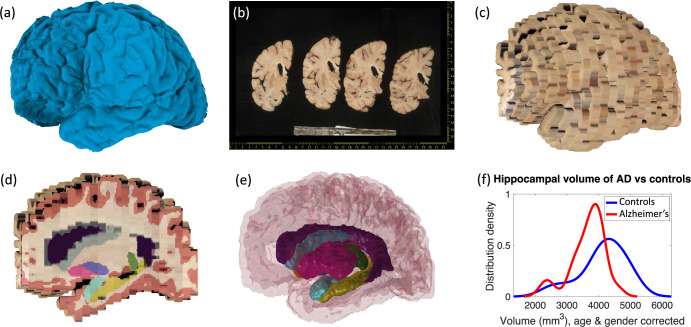
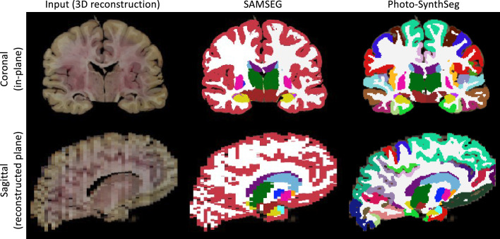
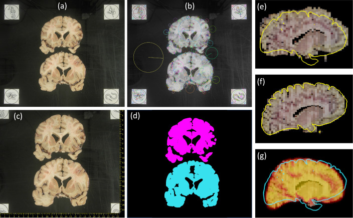
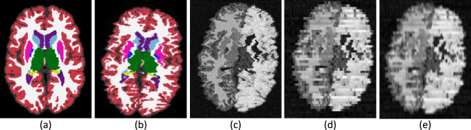
We present open-source tools for three-dimensional (3D) analysis of photographs of dissected slices of human brains, which are routinely acquired in brain banks but seldom used for quantitative analysis. Our tools can: (1) 3D reconstruct a volume from the photographs and, optionally, a surface scan; and (2) produce a high-resolution 3D segmentation into 11 brain regions per hemisphere (22 in total), independently of the slice thickness. Our tools can be used as a substitute for ex vivo magnetic resonance imaging (MRI), which requires access to an MRI scanner, ex vivo scanning expertise, and considerable financial resources. We tested our tools on synthetic and real data from two NIH Alzheimer's Disease Research Centers. The results show that our methodology yields accurate 3D reconstructions, segmentations, and volumetric measurements that are highly correlated to those from MRI. Our method also detects expected differences between confirmed Alzheimer's disease cases and controls. The tools are available in our widespread neuroimaging suite 'FreeSurfer' (https://surfer.nmr.mgh.harvard.edu/fswiki/PhotoTools).

摘要

我们提供了用于分析人脑解剖切片照片的开源工具,这些工具通常在脑库中获取,但很少用于定量分析。我们的工具可以:(1)从照片中(可选地从表面扫描中)重建体积;(2)将高分辨率的 3D 分割为每个半球的 11 个脑区(总共 22 个),而与切片厚度无关。我们的工具可以替代需要访问 MRI 扫描仪、体外扫描专业知识和大量资金的体外磁共振成像(MRI)。我们在来自两个 NIH 阿尔茨海默病研究中心的合成和真实数据上测试了我们的工具。结果表明,我们的方法产生的 3D 重建、分割和体积测量结果与 MRI 非常相关。我们的方法还检测到确认的阿尔茨海默病病例和对照组之间的预期差异。这些工具可在我们广泛使用的神经影像学套件 'FreeSurfer'(https://surfer.nmr.mgh.harvard.edu/fswiki/PhotoTools)中使用。

https://cdn.ncbi.nlm.nih.gov/pmc/blobs/ce6b/11186625/8f5844be87dd/elife-91398-app1-fig3.jpg
https://cdn.ncbi.nlm.nih.gov/pmc/blobs/ce6b/11186625/cb68a7d8c851/elife-91398-fig1.jpg
https://cdn.ncbi.nlm.nih.gov/pmc/blobs/ce6b/11186625/3b0c5a8e9325/elife-91398-fig2.jpg
https://cdn.ncbi.nlm.nih.gov/pmc/blobs/ce6b/11186625/93436b101703/elife-91398-fig3.jpg
https://cdn.ncbi.nlm.nih.gov/pmc/blobs/ce6b/11186625/af2c50c77495/elife-91398-fig4.jpg
https://cdn.ncbi.nlm.nih.gov/pmc/blobs/ce6b/11186625/1e2a692deb58/elife-91398-fig5.jpg
https://cdn.ncbi.nlm.nih.gov/pmc/blobs/ce6b/11186625/01748fe2491f/elife-91398-fig6.jpg
https://cdn.ncbi.nlm.nih.gov/pmc/blobs/ce6b/11186625/77b4a9f40af9/elife-91398-app1-fig1.jpg
https://cdn.ncbi.nlm.nih.gov/pmc/blobs/ce6b/11186625/9c67e97573ea/elife-91398-app1-fig2.jpg
https://cdn.ncbi.nlm.nih.gov/pmc/blobs/ce6b/11186625/8f5844be87dd/elife-91398-app1-fig3.jpg

相似文献

1
Machine learning of dissection photographs and surface scanning for quantitative 3D neuropathology.基于断层照片和表面扫描的机器学习实现定量 3D 神经病理学
Elife. 2024 Jun 19;12:RP91398. doi: 10.7554/eLife.91398.
2
Machine learning of dissection photographs and surface scanning for quantitative 3D neuropathology.用于定量三维神经病理学的解剖照片机器学习与表面扫描
bioRxiv. 2024 Jan 30:2023.06.08.544050. doi: 10.1101/2023.06.08.544050.
3
A ready-to-use machine learning tool for symmetric multi-modality registration of brain MRI.一种用于脑 MRI 对称多模态配准的即用型机器学习工具。
Sci Rep. 2023 Apr 24;13(1):6657. doi: 10.1038/s41598-023-33781-0.
4
Structural magnetic resonance imaging for the early diagnosis of dementia due to Alzheimer's disease in people with mild cognitive impairment.用于轻度认知障碍患者阿尔茨海默病所致痴呆早期诊断的结构磁共振成像
Cochrane Database Syst Rev. 2020 Mar 2;3(3):CD009628. doi: 10.1002/14651858.CD009628.pub2.
5
Determination of Alzheimer's disease based on morphology and atrophy using machine learning combined with automated segmentation.基于机器学习结合自动分割的形态学和萎缩分析诊断阿尔茨海默病。
Acta Radiol. 2024 Apr;65(4):359-366. doi: 10.1177/02841851231218384. Epub 2024 Jan 9.
6
A generalizable brain extraction net (BEN) for multimodal MRI data from rodents, nonhuman primates, and humans.一种可推广的用于啮齿动物、非人灵长类动物和人类多模态 MRI 数据的大脑提取网络(BEN)。
Elife. 2022 Dec 22;11:e81217. doi: 10.7554/eLife.81217.
7
Three-dimensional histology reveals dissociable human hippocampal long-axis gradients of Alzheimer's pathology.三维组织学揭示了可分离的人类海马体阿尔茨海默病病理的长轴梯度。
Alzheimers Dement. 2024 Apr;20(4):2606-2619. doi: 10.1002/alz.13695. Epub 2024 Feb 18.
8
Fast three-dimensional image generation for healthy brain aging using diffeomorphic registration.使用可变形配准快速生成健康大脑老化的三维图像。
Hum Brain Mapp. 2023 Mar;44(4):1289-1308. doi: 10.1002/hbm.26165. Epub 2022 Dec 5.
9
Hippocampal volume change measurement: quantitative assessment of the reproducibility of expert manual outlining and the automated methods FreeSurfer and FIRST.海马体积变化测量:专家手动勾勒以及自动化方法FreeSurfer和FIRST的可重复性定量评估。
Neuroimage. 2014 May 15;92:169-81. doi: 10.1016/j.neuroimage.2014.01.058. Epub 2014 Feb 9.
10
Multi-atlas segmentation of the whole hippocampus and subfields using multiple automatically generated templates.使用多个自动生成的模板对整个海马体及其子区进行多图谱分割。
Neuroimage. 2014 Nov 1;101:494-512. doi: 10.1016/j.neuroimage.2014.04.054. Epub 2014 Apr 29.

引用本文的文献

1
Pseudo-Rendering for Resolution and Topology-Invariant Cortical Parcellation.用于分辨率和拓扑不变皮质分区的伪渲染
Mach Learn Med Imaging. 2025;15242:74-84. doi: 10.1007/978-3-031-73290-4_8. Epub 2024 Oct 23.
2
Synthetic data in generalizable, learning-based neuroimaging.可推广的基于学习的神经影像学中的合成数据。
Imaging Neurosci (Camb). 2024 Nov 19;2:1-22. doi: 10.1162/imag_a_00337. eCollection 2024 Nov 1.
3
Inferotemporal face patches are histo-architectonically distinct.下颞部面孔区在组织形态学上是不同的。

本文引用的文献

1
SynthSeg: Segmentation of brain MRI scans of any contrast and resolution without retraining.SynthSeg:无需重新训练即可对任何对比度和分辨率的脑 MRI 扫描进行分割。
Med Image Anal. 2023 May;86:102789. doi: 10.1016/j.media.2023.102789. Epub 2023 Feb 25.
2
Robust machine learning segmentation for large-scale analysis of heterogeneous clinical brain MRI datasets.用于大规模分析异质临床脑 MRI 数据集的稳健机器学习分割。
Proc Natl Acad Sci U S A. 2023 Feb 28;120(9):e2216399120. doi: 10.1073/pnas.2216399120. Epub 2023 Feb 21.
3
SynthSR: A public AI tool to turn heterogeneous clinical brain scans into high-resolution T1-weighted images for 3D morphometry.
Cell Rep. 2024 Sep 24;43(9):114732. doi: 10.1016/j.celrep.2024.114732. Epub 2024 Sep 12.
SynthSR:一个公共 AI 工具,可将异质临床大脑扫描转换为用于 3D 形态测量的高分辨率 T1 加权图像。
Sci Adv. 2023 Feb 3;9(5):eadd3607. doi: 10.1126/sciadv.add3607. Epub 2023 Feb 1.
4
Protocol for the Systematic Fixation, Circuit-Based Sampling, and Qualitative and Quantitative Neuropathological Analysis of Human Brain Tissue.人类脑组织的系统固定、基于电路的采样以及定性和定量神经病理学分析的方案。
Methods Mol Biol. 2023;2561:3-30. doi: 10.1007/978-1-0716-2655-9_1.
5
Leveraging Neuroimaging Tools to Assess Precision and Accuracy in an Alzheimer's Disease Neuropathologic Sampling Protocol.利用神经影像学工具评估阿尔茨海默病神经病理学采样方案中的精准度和准确性。
Front Neurosci. 2021 Aug 18;15:693242. doi: 10.3389/fnins.2021.693242. eCollection 2021.
6
7 Tesla MRI of the ex vivo human brain at 100 micron resolution.7 特斯拉 MRI 对离体人脑的 100 微米分辨率成像。
Sci Data. 2019 Oct 30;6(1):244. doi: 10.1038/s41597-019-0254-8.
7
A Survey of Methods for 3D Histology Reconstruction.三维组织学重建方法研究综述。
Med Image Anal. 2018 May;46:73-105. doi: 10.1016/j.media.2018.02.004. Epub 2018 Feb 21.
8
Towards a unified analysis of brain maturation and aging across the entire lifespan: A MRI analysis.迈向整个生命周期大脑成熟和衰老的统一分析:一项 MRI 分析。
Hum Brain Mapp. 2017 Nov;38(11):5501-5518. doi: 10.1002/hbm.23743. Epub 2017 Jul 24.
9
Deep Learning for Brain MRI Segmentation: State of the Art and Future Directions.用于脑磁共振成像分割的深度学习:现状与未来方向。
J Digit Imaging. 2017 Aug;30(4):449-459. doi: 10.1007/s10278-017-9983-4.
10
Associations between hippocampal morphometry and neuropathologic markers of Alzheimer's disease using 7 T MRI.使用7T磁共振成像技术研究海马形态测量与阿尔茨海默病神经病理学标志物之间的关联。
Neuroimage Clin. 2017 Apr 21;15:56-61. doi: 10.1016/j.nicl.2017.04.020. eCollection 2017.