Athinoula A. Martinos Center for Biomedical Imaging, Massachusetts General Hospital and Harvard Medical School, Boston, MA, USA.
Centre for Medical Image Computing, Department of Computer Science, University College London, London, UK.
Sci Adv. 2023 Feb 3;9(5):eadd3607. doi: 10.1126/sciadv.add3607. Epub 2023 Feb 1.
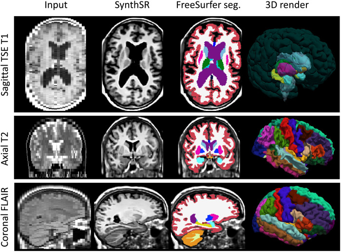
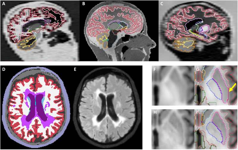
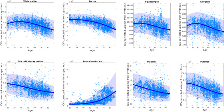
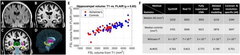
Every year, millions of brain magnetic resonance imaging (MRI) scans are acquired in hospitals across the world. These have the potential to revolutionize our understanding of many neurological diseases, but their morphometric analysis has not yet been possible due to their anisotropic resolution. We present an artificial intelligence technique, "SynthSR," that takes clinical brain MRI scans with any MR contrast (T1, T2, etc.), orientation (axial/coronal/sagittal), and resolution and turns them into high-resolution T1 scans that are usable by virtually all existing human neuroimaging tools. We present results on segmentation, registration, and atlasing of >10,000 scans of controls and patients with brain tumors, strokes, and Alzheimer's disease. SynthSR yields morphometric results that are very highly correlated with what one would have obtained with high-resolution T1 scans. SynthSR allows sample sizes that have the potential to overcome the power limitations of prospective research studies and shed new light on the healthy and diseased human brain.
每年,全球医院都会进行数百万次脑部磁共振成像 (MRI) 扫描。这些扫描有可能彻底改变我们对许多神经疾病的理解,但由于其各向异性分辨率,目前还无法对其进行形态计量分析。我们提出了一种人工智能技术“SynthSR”,它可以将具有任何 MR 对比(T1、T2 等)、方位(轴位/冠状位/矢状位)和分辨率的临床脑部 MRI 扫描转换为高分辨率 T1 扫描,几乎所有现有的人类神经影像学工具都可以使用。我们展示了对 >10000 例脑部肿瘤、中风和阿尔茨海默病患者和对照者的扫描进行分割、配准和图谱绘制的结果。SynthSR 产生的形态计量结果与使用高分辨率 T1 扫描获得的结果非常高度相关。SynthSR 允许增加样本量,有可能克服前瞻性研究的局限性,并为健康和患病的人脑提供新的认识。