Vela-Alcántara Ana Monserrat, Santiago-García Juan, Barragán-Palacios Madeleine, León-Chacón Aylin, Domínguez-Pantoja Marilú, Barceinas-Dávila Irene, Juárez-Aguilar Enrique, Tamariz Elisa
Programa de Doctorado en Ciencias de la Salud, Instituto de Ciencias de la Salud, Universidad Veracruzana, Xalapa, Mexico.
Laboratorio de Cultivo Celular, Departamento de Biomedicina, Instituto de Ciencias de la Salud, Universidad Veracruzana, Xalapa, Mexico.
Front Cell Dev Biol. 2024 Jun 5;12:1352233. doi: 10.3389/fcell.2024.1352233. eCollection 2024.
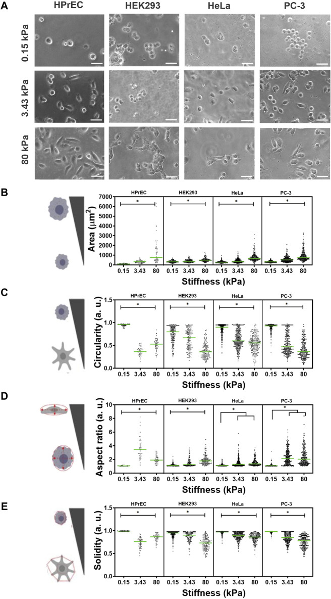
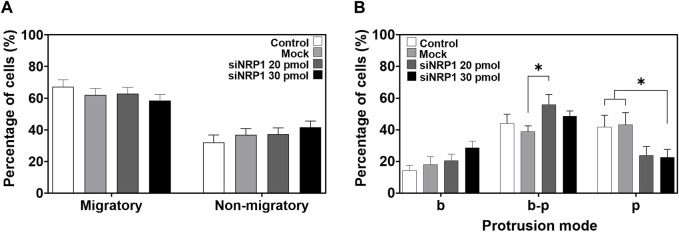
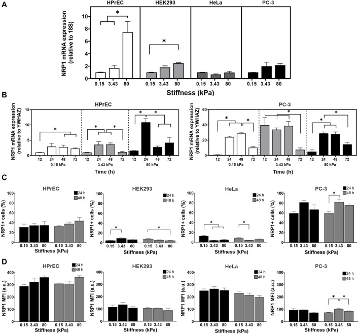
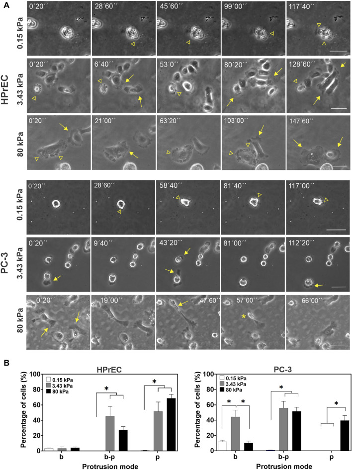
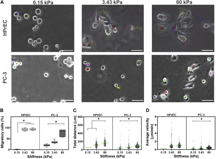
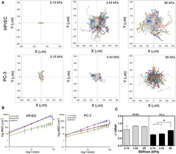
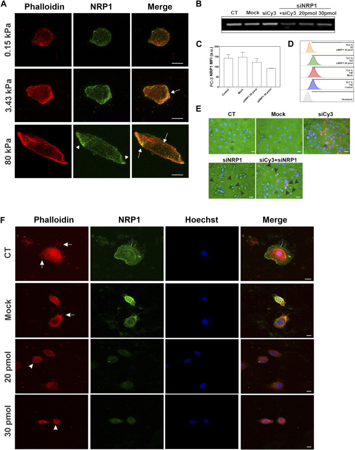
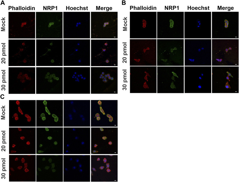
Physical changes in the tumor microenvironment, such as increased stiffness, regulate cancer hallmarks and play an essential role in gene expression, cell morphology, migration, and malignancy. However, the response of cancer cells to stiffness is not homogeneous and varies depending on the cell type and its mechanosensitivity. In this study, we investigated the differential responses of cervical (HeLa) and prostate (PC-3) cancer cell lines, as well as non-tumoral cell lines (HEK293 and HPrEC), to stiffness using polyacrylamide hydrogels mimicking normal and tumoral tissues. We analyzed cell morphology, migration, and the expression of neuropilin 1 (NRP1), a receptor involved in angiogenesis, cell migration, and extracellular matrix remodeling, known to be associated with cancer progression and poor prognosis. Our findings reveal that NRP1 expression increases on substrates mimicking the high stiffness characteristic of tumoral tissue in the non-tumoral cell lines HPrEC and HEK293. Conversely, in tumoral PC-3 cells, stiffness resembling normal prostate tissue induces an earlier and more sustained expression of NRP1. Furthermore, we observed that stiffness influences cell spreading, pseudopodia formation, and the mode of cell protrusion during migration. Soft substrates predominantly trigger bleb cell protrusion, while pseudopodia protrusions increase on substrates mimicking normal and tumor-like stiffnesses in HPrEC cells compared to PC-3 cells. Stiffer substrates also enhance the percentage of migratory cells, as well as their velocity and total displacement, in both non-tumoral and tumoral prostate cells. However, they only improve the persistence of migration in tumoral PC-3 cells. Moreover, we found that NRP1 co-localizes with actin, and its suppression impairs tumoral PC-3 spreading while decreasing pseudopodia protrusion mode. Our results suggest that the modulation of NRP1 expression by the stiffness can be a feedback loop to promote malignancy in non-tumoral and cancer cells, contingent upon the mechanosensitivity of the cells.
肿瘤微环境中的物理变化,如硬度增加,可调节癌症特征,并在基因表达、细胞形态、迁移和恶性肿瘤形成中发挥重要作用。然而,癌细胞对硬度的反应并不一致,而是因细胞类型及其机械敏感性而异。在本研究中,我们使用模拟正常和肿瘤组织的聚丙烯酰胺水凝胶,研究了宫颈癌细胞系(HeLa)和前列腺癌细胞系(PC-3)以及非肿瘤细胞系(HEK293和HPrEC)对硬度的差异反应。我们分析了细胞形态、迁移以及神经纤毛蛋白1(NRP1)的表达,NRP1是一种参与血管生成、细胞迁移和细胞外基质重塑的受体,已知与癌症进展和不良预后相关。我们的研究结果表明,在非肿瘤细胞系HPrEC和HEK293中,模拟肿瘤组织高硬度特征的底物上NRP1表达增加。相反,在肿瘤PC-3细胞中,类似正常前列腺组织的硬度诱导NRP1更早且更持续的表达。此外,我们观察到硬度会影响细胞铺展、伪足形成以及迁移过程中的细胞突起模式。柔软的底物主要触发泡状细胞突起,而与PC-3细胞相比,在模拟正常和肿瘤样硬度的底物上,HPrEC细胞中的伪足突起增加。更硬的底物还会提高非肿瘤和肿瘤前列腺细胞中迁移细胞的百分比、速度和总位移。然而,它们仅改善肿瘤PC-3细胞迁移的持续性。此外,我们发现NRP1与肌动蛋白共定位,其抑制会损害肿瘤PC-3细胞的铺展,同时减少伪足突起模式。我们的结果表明,硬度对NRP1表达的调节可能是促进非肿瘤细胞和癌细胞恶性肿瘤形成的反馈回路,这取决于细胞的机械敏感性。