• 文献检索
  • 文档翻译
  • 深度研究
  • 学术资讯
  • Suppr Zotero 插件Zotero 插件
  • 邀请有礼
  • 套餐&价格
  • 历史记录
应用&插件
Suppr Zotero 插件Zotero 插件浏览器插件Mac 客户端Windows 客户端微信小程序
定价
高级版会员购买积分包购买API积分包
服务
文献检索文档翻译深度研究API 文档MCP 服务
关于我们
关于 Suppr公司介绍联系我们用户协议隐私条款
关注我们

Suppr 超能文献

核心技术专利:CN118964589B侵权必究
粤ICP备2023148730 号-1Suppr @ 2026

文献检索

告别复杂PubMed语法,用中文像聊天一样搜索,搜遍4000万医学文献。AI智能推荐,让科研检索更轻松。

立即免费搜索

文件翻译

保留排版,准确专业,支持PDF/Word/PPT等文件格式,支持 12+语言互译。

免费翻译文档

深度研究

AI帮你快速写综述,25分钟生成高质量综述,智能提取关键信息,辅助科研写作。

立即免费体验

胆囊切除术后并发症:放射科医生需要了解的内容。

Post-operative complications of cholecystectomy: what the radiologist needs to know.

作者信息

Kazi Irfan Amir, Siddiqui M Azfar, Thimmappa Nanda Deepa, Abdelaziz Amr, Gaballah Ayman H, Davis Ryan, Kimchi Eric, Hammoud Ghassan, Syed Kazi A, Nasrullah Ayesha

机构信息

Department of Radiology, University Hospital, University of Missouri, 1 Hospital Drive, Columbia, MO, 65212, USA.

Department of Radiology, University of Missouri, Columbia, MO, USA.

出版信息

Abdom Radiol (NY). 2025 Jan;50(1):109-130. doi: 10.1007/s00261-024-04387-5. Epub 2024 Jun 28.

DOI:10.1007/s00261-024-04387-5
PMID:38940909
原文链接:https://pmc.ncbi.nlm.nih.gov/articles/PMC11711778/
Abstract

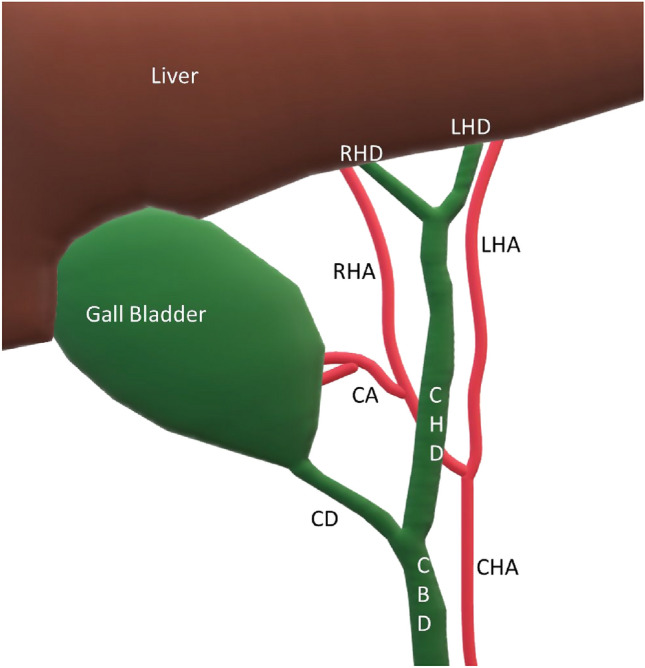
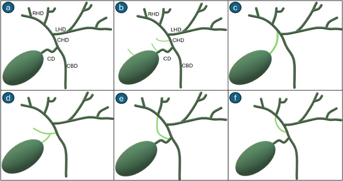
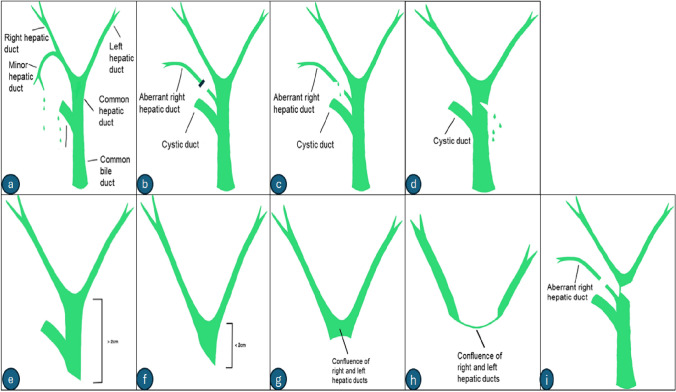
Cholecystectomy is one of the most performed surgical procedures. The safety of this surgery notwithstanding, the sheer volume of operations results in a notable incidence of post-cholecystectomy complications. Early and accurate diagnosis of such complications is essential for timely and effective management. Imaging techniques are critical for this purpose, aiding in distinguishing between expected postsurgical changes and true complications. This review highlights current knowledge on the indications for cholecystectomy, pertinent surgical anatomy and surgical technique, and the recognition of anatomical variants that may complicate surgery. The article also outlines the roles of various imaging modalities in identifying complications, the spectrum of possible postsurgical anatomical changes, and the implications of such findings. Furthermore, we explore the array of complications that can arise post-cholecystectomy, such as biliary system injuries, gallstone-related issues, vascular complications, and the formation of postsurgical collections. Radiologists should be adept at identifying normal and abnormal postoperative findings to guide patient management effectively.

摘要

胆囊切除术是最常施行的外科手术之一。尽管该手术具有安全性,但手术量巨大导致胆囊切除术后并发症的发生率显著。对此类并发症进行早期准确诊断对于及时有效的管理至关重要。成像技术对于此目的至关重要,有助于区分预期的术后变化和真正的并发症。本综述重点介绍了关于胆囊切除术的适应证、相关手术解剖结构和手术技术,以及识别可能使手术复杂化的解剖变异的当前知识。本文还概述了各种成像方式在识别并发症、术后可能的解剖变化范围以及此类发现的意义方面的作用。此外,我们探讨了胆囊切除术后可能出现的一系列并发症,如胆道系统损伤、胆结石相关问题、血管并发症以及术后积液的形成。放射科医生应善于识别术后正常和异常表现,以有效地指导患者管理。

https://cdn.ncbi.nlm.nih.gov/pmc/blobs/40fe/11711778/0c0a87975e38/261_2024_4387_Fig22_HTML.jpg
https://cdn.ncbi.nlm.nih.gov/pmc/blobs/40fe/11711778/b8ed6e53839d/261_2024_4387_Fig1_HTML.jpg
https://cdn.ncbi.nlm.nih.gov/pmc/blobs/40fe/11711778/ea57c0303469/261_2024_4387_Fig2_HTML.jpg
https://cdn.ncbi.nlm.nih.gov/pmc/blobs/40fe/11711778/2d8f2422b683/261_2024_4387_Fig3_HTML.jpg
https://cdn.ncbi.nlm.nih.gov/pmc/blobs/40fe/11711778/6b97c5fba0af/261_2024_4387_Fig4_HTML.jpg
https://cdn.ncbi.nlm.nih.gov/pmc/blobs/40fe/11711778/4acef04b3ec0/261_2024_4387_Fig5_HTML.jpg
https://cdn.ncbi.nlm.nih.gov/pmc/blobs/40fe/11711778/eb254b9d518b/261_2024_4387_Fig6_HTML.jpg
https://cdn.ncbi.nlm.nih.gov/pmc/blobs/40fe/11711778/54445a4404f8/261_2024_4387_Fig7_HTML.jpg
https://cdn.ncbi.nlm.nih.gov/pmc/blobs/40fe/11711778/73c4f3edcc4c/261_2024_4387_Fig8_HTML.jpg
https://cdn.ncbi.nlm.nih.gov/pmc/blobs/40fe/11711778/cf5b17cfaf7e/261_2024_4387_Fig9_HTML.jpg
https://cdn.ncbi.nlm.nih.gov/pmc/blobs/40fe/11711778/bbc4460b974e/261_2024_4387_Fig10_HTML.jpg
https://cdn.ncbi.nlm.nih.gov/pmc/blobs/40fe/11711778/c01566ccb139/261_2024_4387_Fig11_HTML.jpg
https://cdn.ncbi.nlm.nih.gov/pmc/blobs/40fe/11711778/a7faf684023d/261_2024_4387_Fig12_HTML.jpg
https://cdn.ncbi.nlm.nih.gov/pmc/blobs/40fe/11711778/904af5615a81/261_2024_4387_Fig13_HTML.jpg
https://cdn.ncbi.nlm.nih.gov/pmc/blobs/40fe/11711778/1bd691d1b94b/261_2024_4387_Fig14_HTML.jpg
https://cdn.ncbi.nlm.nih.gov/pmc/blobs/40fe/11711778/03d0723198bb/261_2024_4387_Fig15_HTML.jpg
https://cdn.ncbi.nlm.nih.gov/pmc/blobs/40fe/11711778/6c45325a4133/261_2024_4387_Fig16_HTML.jpg
https://cdn.ncbi.nlm.nih.gov/pmc/blobs/40fe/11711778/29da8fc3eb4c/261_2024_4387_Fig17_HTML.jpg
https://cdn.ncbi.nlm.nih.gov/pmc/blobs/40fe/11711778/1d40010af2d2/261_2024_4387_Fig18_HTML.jpg
https://cdn.ncbi.nlm.nih.gov/pmc/blobs/40fe/11711778/76e5dd0a0630/261_2024_4387_Fig19_HTML.jpg
https://cdn.ncbi.nlm.nih.gov/pmc/blobs/40fe/11711778/3ec5fe65ebac/261_2024_4387_Fig20_HTML.jpg
https://cdn.ncbi.nlm.nih.gov/pmc/blobs/40fe/11711778/b6980934f07d/261_2024_4387_Fig21_HTML.jpg
https://cdn.ncbi.nlm.nih.gov/pmc/blobs/40fe/11711778/0c0a87975e38/261_2024_4387_Fig22_HTML.jpg

相似文献

1
Post-operative complications of cholecystectomy: what the radiologist needs to know.胆囊切除术后并发症:放射科医生需要了解的内容。
Abdom Radiol (NY). 2025 Jan;50(1):109-130. doi: 10.1007/s00261-024-04387-5. Epub 2024 Jun 28.
2
Do not get stumped: multimodality imaging findings of early and late post-cholecystectomy complications.别被难住了:胆囊切除术后早晚期并发症的多模态影像学表现。
Emerg Radiol. 2023 Jun;30(3):351-362. doi: 10.1007/s10140-023-02131-y. Epub 2023 Apr 12.
3
Comprehensive Imaging Insights into Post-Cholecystectomy Complications for Enhanced Clinical Practice.胆囊切除术后并发症的综合影像学见解以加强临床实践
Clin Med Res. 2024 Dec;22(4):206-214. doi: 10.3121/cmr.2025.1985.
4
Endoscopic management of postoperative bile leaks.术后胆漏的内镜治疗
Hepatobiliary Pancreat Dis Int. 2006 May;5(2):273-7.
5
MRI evaluation of bile duct injuries and other post-cholecystectomy complications.MRI 评估胆管损伤和其他胆囊切除术后并发症。
Abdom Radiol (NY). 2021 Jul;46(7):3086-3104. doi: 10.1007/s00261-020-02947-z. Epub 2021 Feb 12.
6
Management of post cholecystectomy vascular injuries.胆囊切除术后血管损伤的处理。
Surgeon. 2019 Dec;17(6):326-333. doi: 10.1016/j.surge.2018.10.002. Epub 2018 Nov 3.
7
Imaging in laparoscopic cholecystectomy--what a radiologist needs to know.
Eur J Radiol. 2014 Jun;83(6):867-879. doi: 10.1016/j.ejrad.2014.02.016. Epub 2014 Mar 3.
8
Biliary complications of surgical procedures: what the radiologist needs to know.手术的胆道并发症:放射科医生需要了解的内容。
Abdom Radiol (NY). 2024 Dec 30. doi: 10.1007/s00261-024-04754-2.
9
The role of ERCP in patients after laparoscopic cholecystectomy.内镜逆行胰胆管造影术在腹腔镜胆囊切除术后患者中的作用。
Am J Gastroenterol. 1994 Sep;89(9):1523-7.
10
Biliary tract injuries during laparoscopic cholecystectomy: three case reports and literature review.腹腔镜胆囊切除术中的胆道损伤:三例报告及文献综述
G Chir. 2010 Jan-Feb;31(1-2):16-9.

引用本文的文献

1
Anatomical variations of hepatic arteries: implications for clinical practice.肝动脉的解剖变异:对临床实践的影响。
Front Surg. 2025 Jun 18;12:1593800. doi: 10.3389/fsurg.2025.1593800. eCollection 2025.
2
Endoscopic management of post-cholecystectomy cystic duct stump biliary leakage: Single-centre experience.胆囊切除术后胆囊管残端胆漏的内镜治疗:单中心经验
Turk J Surg. 2025 May 30;41(2):193-197. doi: 10.47717/turkjsurg.2025.6616.
3
Minimally Invasive Approaches for High-Risk and Elderly Patients With Acute Cholecystitis: A Systematic Review of Techniques and Outcomes.

本文引用的文献

1
Imaging after Pancreatic Surgery: Expected Findings and Postoperative Complications.胰腺手术后的影像学检查:预期结果与术后并发症
Radiographics. 2024 Jan;44(1):e230061. doi: 10.1148/rg.230061.
2
A retrospective study of laparoscopic, robotic-assisted, and open emergent/urgent cholecystectomy based on the PINC AI Healthcare Database 2017-2020.基于 PINC AI Healthcare Database 2017-2020 的腹腔镜、机器人辅助和开放性紧急/紧急胆囊切除术的回顾性研究。
World J Emerg Surg. 2023 Nov 30;18(1):55. doi: 10.1186/s13017-023-00521-8.
3
Vascular injury during laparoscopic cholecystectomy: An often-overlooked complication.
高危及老年急性胆囊炎患者的微创治疗方法:技术与疗效的系统评价
Cureus. 2025 Jan 30;17(1):e78271. doi: 10.7759/cureus.78271. eCollection 2025 Jan.
腹腔镜胆囊切除术中的血管损伤:一种常被忽视的并发症。
World J Gastrointest Surg. 2023 Mar 27;15(3):338-345. doi: 10.4240/wjgs.v15.i3.338.
4
Computed tomography-guided percutaneous cholecystostomy: a single institution's 6-year experience.计算机断层扫描引导下经皮胆囊造瘘术:单机构6年经验
Ann Gastroenterol. 2022 Nov-Dec;35(6):668-672. doi: 10.20524/aog.2022.0755. Epub 2022 Oct 17.
5
Consequences of the spilled gallstones during laparoscopic cholecystectomy: a systematic review.腹腔镜胆囊切除术中胆囊结石溢出的后果:系统评价。
World J Emerg Surg. 2022 Nov 2;17(1):57. doi: 10.1186/s13017-022-00456-6.
6
Multimodality Imaging of Cholecystectomy Complications.胆囊切除术并发症的多模态影像学
Radiographics. 2022 Sep-Oct;42(5):1303-1319. doi: 10.1148/rg.210106. Epub 2022 Jul 29.
7
RISK FACTORS FOR SURGICAL WOUND INFECTION AFTER ELECTIVE LAPAROSCOPIC CHOLECYSTECTOMY.择期腹腔镜胆囊切除术术后手术部位感染的危险因素。
Arq Bras Cir Dig. 2022 Jun 17;35:e1655. doi: 10.1590/0102-672020210002e1655. eCollection 2022.
8
Multimodality imaging of Surgicel, an important mimic of post-operative complication in the thorax.Surgicel的多模态成像,其是胸部术后并发症的重要模拟物。
BJR Open. 2021 Nov 24;3(1):20210031. doi: 10.1259/bjro.20210031. eCollection 2021.
9
2020 WSES guidelines for the detection and management of bile duct injury during cholecystectomy.2020 WSES 指南:胆囊切除术胆道损伤的检测与处理。
World J Emerg Surg. 2021 Jun 10;16(1):30. doi: 10.1186/s13017-021-00369-w.
10
MRI evaluation of bile duct injuries and other post-cholecystectomy complications.MRI 评估胆管损伤和其他胆囊切除术后并发症。
Abdom Radiol (NY). 2021 Jul;46(7):3086-3104. doi: 10.1007/s00261-020-02947-z. Epub 2021 Feb 12.