School of Dentistry, Shaheed Zulfiqar Ali Bhutto Medical University, Islamabad, Pakistan.
Department of Orthodontics, Islamic International Dental College, Riphah International University, Islamabad, Pakistan.
PeerJ. 2024 Jun 28;12:e17645. doi: 10.7717/peerj.17645. eCollection 2024.
The aim of this study was threefold. Firstly, it aimed to introduce and detail a novel method for chemically etching the bases of stainless-steel orthodontic brackets. Secondly, the study sought to investigate the structural alterations within the brackets' microstructure following chemical etching compared to those with sandblasted bases, using electron microscopy analysis. Lastly, the study aimed to evaluate and compare the long-term durability and survivability of orthodontic brackets with chemically etched bases those with sandblasted bases, both bonded using the conventional acid etch technique with Transbond XT adhesive, over an 18-month follow-up period.
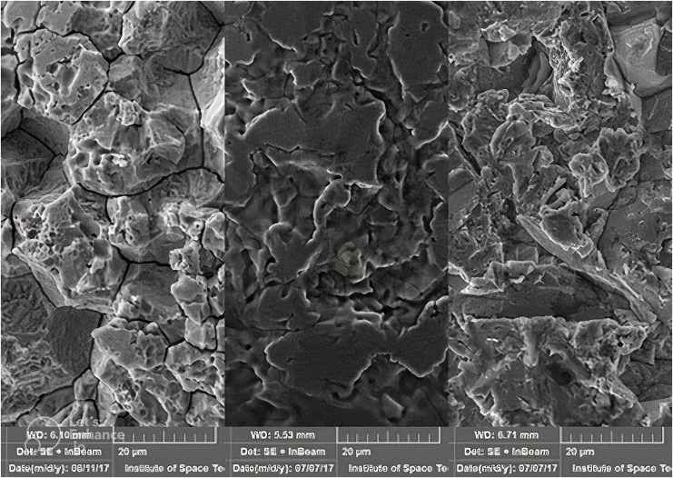
The study was a randomized clinical control trial with triple blinding and split-mouth study design and consisted of two groups. The brackets in the sandblasted group were prepared by sandblasting the intaglio surface of the base of the bracket with 50 µm SiO particles. Hydrofluoric acid was used to roughen the base in the acid-etched group. The bases of the brackets were viewed under an electron microscope to analyze the topographical changes.
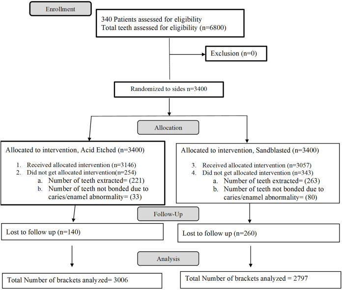
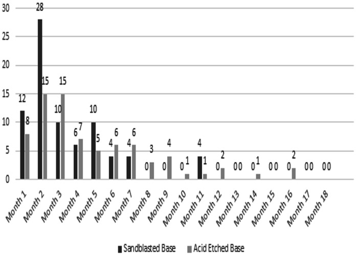
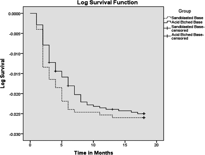
A total of 5,803 brackets (3,006 acid-etch, 2,797 sandblasted) in 310 patients were bonded, in a split-mouth design by the same operator. The patients were followed for 18 months. The failure rate of 2.59% and 2.7% was noted in an acid-etched and sandblasted group, respectively. There was a close approximation of curves in the Kaplan-Meier plot, and the survival distribution of the two groups in the log-rank (Mantel-Cox) test was insignificant; x2 = 0.062 ( value = 0.804).
Acid etching if the bases of the brackets can be used as an alternative to sandblasting furthermore acid etching can be performed on the chair side.
本研究旨在实现三个目标。首先,引入并详细介绍一种新的不锈钢正畸托槽基底化学蚀刻方法。其次,通过电子显微镜分析,研究比较化学蚀刻与喷砂基底托槽后,其微观结构的变化。最后,评估并比较使用传统酸蚀技术和 Transbond XT 粘结剂粘结,经化学蚀刻基底和喷砂基底的正畸托槽在 18 个月随访期间的长期耐用性和存活率。
该研究为随机临床对照试验,采用三重盲法和分面研究设计,共包括两组。喷砂组通过用 50µmSiO 粒子喷砂托槽基底的凹面来制备托槽。酸蚀组使用氢氟酸使基底粗糙化。通过电子显微镜观察托槽基底,分析形貌变化。
共粘结 5803 个托槽(3006 个酸蚀,2797 个喷砂),由同一位操作人员按分面设计粘结于 310 名患者。患者随访 18 个月。酸蚀组和喷砂组的失败率分别为 2.59%和 2.7%。Kaplan-Meier 绘图中曲线接近,对数秩(Mantel-Cox)检验中两组的生存分布无显著差异;x2=0.062( 值=0.804)。
酸蚀可作为替代喷砂的方法,此外酸蚀可在椅旁进行。