Meliti Abdelrazak, Alardati Hosam, Khayat Manal, Alruqi Abdullah
Department of Pathology and Laboratory Medicine, King Faisal Specialist Hospital and Research Centre, Jeddah, SAU.
Department of Pathology and Laboratory Medicine, King Abdulaziz Medical City, Jeddah, SAU.
Cureus. 2024 Jun 8;16(6):e61940. doi: 10.7759/cureus.61940. eCollection 2024 Jun.
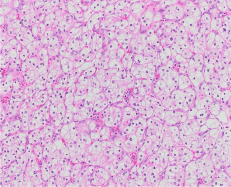
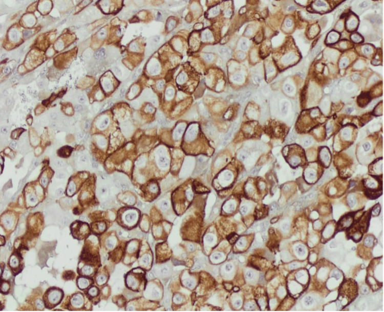
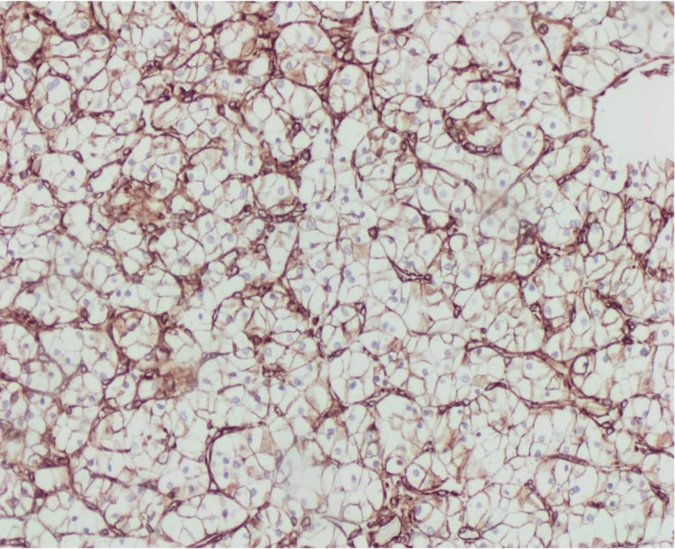
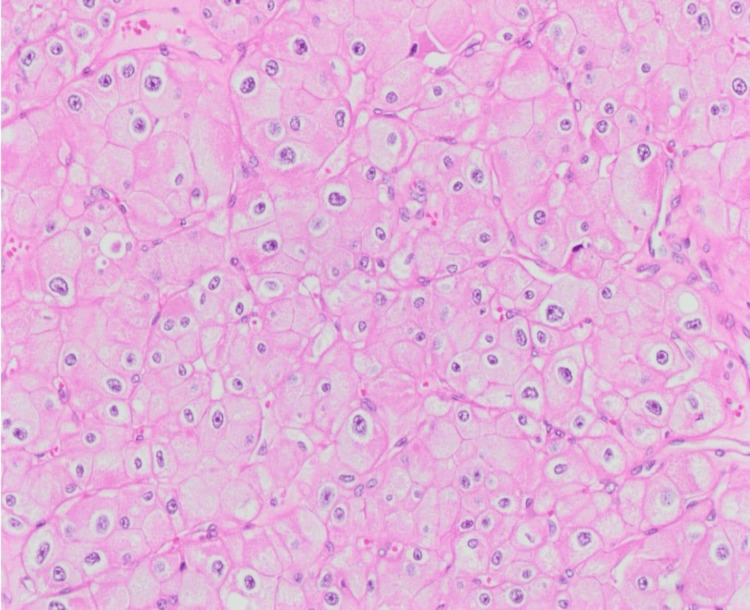
Renal cell carcinoma (RCC) is a diverse array of cancers arising from renal tubular epithelial cells. RCC presenting with distinct morphological subtypes, such as the simultaneous presence of chromophobe RCC (chRCC) and clear cell RCC (ccRCC) lesions within the same kidney, is rare. We present the case of a 79-year-old female with a history of breast cancer who presented to our facility with right flank pain. Further investigations using CT of the abdomen and pelvis revealed a Bosniak type 4 cyst with a mural nodule in the right kidney. Furthermore, another well-defined, solid lesion measuring 2.8 × 2.6 cm was observed in the same area. The patient underwent a right radical nephrectomy. The macroscopic examination of the kidney revealed the presence of three cysts, with the largest measuring up to 7.5 cm. Moreover, a distinctly demarcated, golden-yellow, solid mass was discerned in the superior pole of the kidney. The mass showed a heterogeneous cut surface with solid and cystic components, measuring 2.8 × 2.6 × 2.0 cm. A less extensive but well-defined, uniform tan mass was also identified within the wall of the largest cyst, which measured 1.2 × 1.0 × 0.7 cm. At this point, the diagnosis of ccRCC and chRCC was established.
肾细胞癌(RCC)是起源于肾小管上皮细胞的多种癌症。肾细胞癌呈现出不同的形态学亚型,例如在同一肾脏内同时存在嫌色性肾细胞癌(chRCC)和透明细胞肾细胞癌(ccRCC)病变的情况较为罕见。我们报告了一例79岁有乳腺癌病史的女性患者,她因右侧胁腹疼痛前来我院就诊。使用腹部和盆腔CT进行的进一步检查显示右肾有一个博斯尼亚克4型囊肿,伴有壁结节。此外,在同一区域观察到另一个边界清晰的实性病变,大小为2.8×2.6厘米。患者接受了右侧根治性肾切除术。对肾脏的宏观检查发现有三个囊肿,最大的囊肿直径达7.5厘米。此外,在肾的上极发现一个界限分明的金黄色实性肿块。该肿块切面呈异质性,有实性和囊性成分,大小为2.8×2.6×2.0厘米。在最大囊肿的壁内还发现一个范围较小但边界清晰、均匀的黄褐色肿块,大小为1.2×1.0×0.7厘米。此时,确诊为透明细胞肾细胞癌和嫌色性肾细胞癌。