• 文献检索
  • 文档翻译
  • 深度研究
  • 学术资讯
  • Suppr Zotero 插件Zotero 插件
  • 邀请有礼
  • 套餐&价格
  • 历史记录
应用&插件
Suppr Zotero 插件Zotero 插件浏览器插件Mac 客户端Windows 客户端微信小程序
定价
高级版会员购买积分包购买API积分包
服务
文献检索文档翻译深度研究API 文档MCP 服务
关于我们
关于 Suppr公司介绍联系我们用户协议隐私条款
关注我们

Suppr 超能文献

核心技术专利:CN118964589B侵权必究
粤ICP备2023148730 号-1Suppr @ 2026

文献检索

告别复杂PubMed语法,用中文像聊天一样搜索,搜遍4000万医学文献。AI智能推荐,让科研检索更轻松。

立即免费搜索

文件翻译

保留排版,准确专业,支持PDF/Word/PPT等文件格式,支持 12+语言互译。

免费翻译文档

深度研究

AI帮你快速写综述,25分钟生成高质量综述,智能提取关键信息,辅助科研写作。

立即免费体验

非小细胞肺癌中尽管主要组织相容性复合体I类严重缺失,但自然杀伤细胞和CD8 T细胞的空间共定位及联合生存获益

Spatial colocalization and combined survival benefit of natural killer and CD8 T cells despite profound MHC class I loss in non-small cell lung cancer.

作者信息

Wessel Remziye E, Ageeb Nardin, Obeid Joseph M, Mauldin Ileana, Goundry Kate A, Hanson Gabriel F, Hossain Mahdin, Lehman Chad, Gentzler Ryan D, Wages Nolan A, Slingluff Craig L, Bullock Timothy N J, Dolatshahi Sepideh, Brown Michael G

机构信息

Department of Biomedical Engineering, University of Virginia (UVA) School of Medicine, Charlottesville, Virginia 22908.

Department of Biology, UVA, Charlottesville, Virginia 22908.

出版信息

bioRxiv. 2024 Jun 25:2024.02.20.581048. doi: 10.1101/2024.02.20.581048.

DOI:10.1101/2024.02.20.581048
PMID:38979183
原文链接:https://pmc.ncbi.nlm.nih.gov/articles/PMC11230195/
Abstract

BACKGROUND

MHC class I (MHC-I) loss is frequent in non-small cell lung cancer (NSCLC) rendering tumor cells resistant to T cell lysis. NK cells kill MHC-I-deficient tumor cells, and although previous work indicated their presence at NSCLC margins, they were functionally impaired. Within, we evaluated whether NK cell and CD8 T cell infiltration and activation vary with MHC-I expression.

METHODS

We used single-stain immunohistochemistry (IHC) and Kaplan-Meier analysis to test the effect of NK cell and CD8 T cell infiltration on overall and disease-free survival. To delineate immune covariates of MHC-I-disparate lung cancers, we used multiplexed immunofluorescence (mIF) imaging followed by multivariate statistical modeling. To identify differences in infiltration and intercellular communication between IFNγ-activated and non-activated lymphocytes, we developed a computational pipeline to enumerate single cell neighborhoods from mIF images followed by multivariate discriminant analysis.

RESULTS

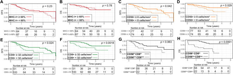
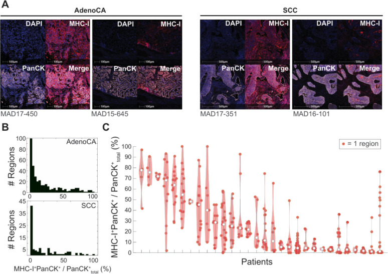
Spatial quantitation of tumor cell MHC-I expression revealed intra- and inter-tumoral heterogeneity, which was associated with the local lymphocyte landscape. IHC analysis revealed that high CD56 cell numbers in patient tumors were positively associated with disease-free survival (DFS) (HR=0.58, =0.064) and overall survival (OS) (HR=0.496, =0.041). The OS association strengthened with high counts of both CD56 and CD8 cells (HR=0.199, <1×10). mIF imaging and multivariate discriminant analysis revealed enrichment of both CD3CD8 T cells and CD3CD56 NK cells in MHC-I-bearing tumors (p<0.05). To infer associations of functional cell states and local cell-cell communication, we analyzed spatial single cell neighborhood profiles to delineate the cellular environments of IFNγ NK cells and T cells. We discovered that both IFNγ NK and CD8 T cells were more frequently associated with other IFNγ lymphocytes in comparison to IFNγ NK cells and CD8 T cells (p<1×10). Moreover, IFNγ lymphocytes were most often found clustered near MHC-I tumor cells.

CONCLUSIONS

Tumor-infiltrating NK cells and CD8 T cells jointly affected control of NSCLC tumor progression. Co-association of NK and CD8 T cells was most evident in MHC-I-bearing tumors, especially in the presence of IFNγ. Frequent co-localization of IFNγ NK cells with other IFNγ lymphocytes in near-neighbor analysis suggests NSCLC lymphocyte activation is coordinately regulated.

摘要

背景

MHC I类分子(MHC-I)缺失在非小细胞肺癌(NSCLC)中很常见,使肿瘤细胞对T细胞裂解产生抗性。自然杀伤细胞(NK细胞)可杀伤缺乏MHC-I的肿瘤细胞,尽管先前的研究表明它们存在于NSCLC边缘,但功能受损。在此,我们评估了NK细胞和CD8 T细胞浸润及激活是否随MHC-I表达而变化。

方法

我们使用单染色免疫组织化学(IHC)和Kaplan-Meier分析来测试NK细胞和CD8 T细胞浸润对总生存期和无病生存期的影响。为了描绘MHC-I不同的肺癌的免疫协变量,我们使用多重免疫荧光(mIF)成像,然后进行多变量统计建模。为了识别IFNγ激活的淋巴细胞和未激活的淋巴细胞在浸润和细胞间通讯方面的差异,我们开发了一个计算流程,从mIF图像中枚举单细胞邻域,然后进行多变量判别分析。

结果

肿瘤细胞MHC-I表达的空间定量揭示了肿瘤内和肿瘤间的异质性,这与局部淋巴细胞格局相关。IHC分析显示,患者肿瘤中高数量的CD56细胞与无病生存期(DFS)呈正相关(HR=0.58,P=0.064)和总生存期(OS)呈正相关(HR=0.496,P=0.041)。当CD56和CD8细胞数量都很高时,OS相关性增强(HR=0.199,P<1×10)。mIF成像和多变量判别分析显示,在表达MHC-I的肿瘤中,CD3+CD8+T细胞和CD3+CD56+NK细胞均富集(P<0.05)。为了推断功能细胞状态与局部细胞间通讯的关联,我们分析了空间单细胞邻域图谱,以描绘IFNγ+NK细胞和T细胞的细胞环境。我们发现,与IFNγ-NK细胞和CD8-T细胞相比,IFNγ+NK细胞和CD8+T细胞与其他IFNγ+淋巴细胞的关联更频繁(P<1×10)。此外,IFNγ+淋巴细胞最常聚集在MHC-I肿瘤细胞附近。

结论

肿瘤浸润性NK细胞和CD8 T细胞共同影响NSCLC肿瘤进展的控制。NK细胞和CD8 T细胞的共关联在表达MHC-I的肿瘤中最为明显,尤其是在存在IFNγ的情况下。在近邻分析中,IFNγ+NK细胞与其他IFNγ+淋巴细胞频繁共定位,表明NSCLC淋巴细胞激活受到协调调节。

https://cdn.ncbi.nlm.nih.gov/pmc/blobs/249b/11230195/0b91b4f01fb9/nihpp-2024.02.20.581048v2-f0006.jpg
https://cdn.ncbi.nlm.nih.gov/pmc/blobs/249b/11230195/08d329a4365e/nihpp-2024.02.20.581048v2-f0001.jpg
https://cdn.ncbi.nlm.nih.gov/pmc/blobs/249b/11230195/20710a0913b0/nihpp-2024.02.20.581048v2-f0002.jpg
https://cdn.ncbi.nlm.nih.gov/pmc/blobs/249b/11230195/3096c75b75f5/nihpp-2024.02.20.581048v2-f0003.jpg
https://cdn.ncbi.nlm.nih.gov/pmc/blobs/249b/11230195/c45f51897978/nihpp-2024.02.20.581048v2-f0004.jpg
https://cdn.ncbi.nlm.nih.gov/pmc/blobs/249b/11230195/35cb7482188d/nihpp-2024.02.20.581048v2-f0005.jpg
https://cdn.ncbi.nlm.nih.gov/pmc/blobs/249b/11230195/0b91b4f01fb9/nihpp-2024.02.20.581048v2-f0006.jpg

相似文献

1
Spatial colocalization and combined survival benefit of natural killer and CD8 T cells despite profound MHC class I loss in non-small cell lung cancer.非小细胞肺癌中尽管主要组织相容性复合体I类严重缺失,但自然杀伤细胞和CD8 T细胞的空间共定位及联合生存获益
bioRxiv. 2024 Jun 25:2024.02.20.581048. doi: 10.1101/2024.02.20.581048.
2
Spatial colocalization and combined survival benefit of natural killer and CD8 T cells despite profound MHC class I loss in non-small cell lung cancer.尽管非小细胞肺癌中 MHC I 类分子严重缺失,但自然杀伤细胞和 CD8 T 细胞的空间共存和联合生存获益。
J Immunother Cancer. 2024 Sep 18;12(9):e009126. doi: 10.1136/jitc-2024-009126.
3
Natural killer cells infiltrating colorectal cancer and MHC class I expression.浸润结直肠癌的自然杀伤细胞与MHC I类分子表达
Mol Immunol. 2005 Feb;42(4):541-6. doi: 10.1016/j.molimm.2004.07.039.
4
Intratumoral NKp46 natural killer cells are spatially distanced from T and MHC-I cells with prognostic implications in soft tissue sarcoma.肿瘤内 NKp46 自然杀伤细胞与 T 细胞和 MHC-I 细胞在空间上分离,这与软组织肉瘤的预后有关。
Front Immunol. 2023 Jul 21;14:1230534. doi: 10.3389/fimmu.2023.1230534. eCollection 2023.
5
NK cells and T cells cooperate during the clinical course of colorectal cancer.自然杀伤细胞和 T 细胞在结直肠癌的临床病程中协同作用。
Oncoimmunology. 2014 Aug 3;3(8):e952197. doi: 10.4161/21624011.2014.952197. eCollection 2014.
6
Adoptive transfer of natural killer cells promotes the anti-tumor efficacy of T cells.自然杀伤细胞的过继性转移可增强T细胞的抗肿瘤功效。
Clin Immunol. 2017 Apr;177:76-86. doi: 10.1016/j.clim.2016.06.013. Epub 2016 Jul 1.
7
Interferon Gamma Induces Changes in Natural Killer (NK) Cell Ligand Expression and Alters NK Cell-Mediated Lysis of Pediatric Cancer Cell Lines.γ干扰素诱导自然杀伤(NK)细胞配体表达的变化并改变NK细胞介导的对小儿癌细胞系的杀伤作用。
Front Immunol. 2017 Apr 6;8:391. doi: 10.3389/fimmu.2017.00391. eCollection 2017.
8
Tumor immune microenvironment analysis of non-small cell lung cancer development through multiplex immunofluorescence.通过多重免疫荧光对非小细胞肺癌发展进行肿瘤免疫微环境分析。
Transl Lung Cancer Res. 2024 Sep 30;13(9):2395-2410. doi: 10.21037/tlcr-24-379. Epub 2024 Sep 20.
9
Proliferating CD8 T Cell Infiltrates Are Associated with Improved Survival in Glioblastoma.增殖的 CD8+T 细胞浸润与胶质母细胞瘤患者的生存改善相关。
Cells. 2021 Dec 1;10(12):3378. doi: 10.3390/cells10123378.
10
[Correlation between Immune Microenvironment Features and EGFR Mutation Status 
in Lung Adenocarcinoma].[肺腺癌免疫微环境特征与表皮生长因子受体突变状态的相关性]
Zhongguo Fei Ai Za Zhi. 2023 Mar 20;26(3):204-216. doi: 10.3779/j.issn.1009-3419.2023.101.07.

本文引用的文献

1
Intratumoral NKp46 natural killer cells are spatially distanced from T and MHC-I cells with prognostic implications in soft tissue sarcoma.肿瘤内 NKp46 自然杀伤细胞与 T 细胞和 MHC-I 细胞在空间上分离,这与软组织肉瘤的预后有关。
Front Immunol. 2023 Jul 21;14:1230534. doi: 10.3389/fimmu.2023.1230534. eCollection 2023.
2
Designing Cancer Immunotherapies That Engage T Cells and NK Cells.设计能同时激活 T 细胞和自然杀伤细胞的癌症免疫疗法。
Annu Rev Immunol. 2023 Apr 26;41:17-38. doi: 10.1146/annurev-immunol-101921-044122. Epub 2022 Nov 29.
3
Altered Maternal Antibody Profiles in Women With Human Immunodeficiency Virus Drive Changes in Transplacental Antibody Transfer.
母体抗体谱的改变会影响 HIV 感染者的胎盘抗体转移。
Clin Infect Dis. 2022 Oct 12;75(8):1359-1369. doi: 10.1093/cid/ciac156.
4
Cancer statistics, 2022.癌症统计数据,2022 年。
CA Cancer J Clin. 2022 Jan;72(1):7-33. doi: 10.3322/caac.21708. Epub 2022 Jan 12.
5
Distinct tumor-infiltrating lymphocyte landscapes are associated with clinical outcomes in localized non-small-cell lung cancer.不同的肿瘤浸润淋巴细胞图谱与局限性非小细胞肺癌的临床结局相关。
Ann Oncol. 2022 Jan;33(1):42-56. doi: 10.1016/j.annonc.2021.09.021. Epub 2021 Oct 13.
6
Antibody Subclass and Glycosylation Shift Following Effective TB Treatment.有效抗结核治疗后抗体亚类和糖基化转移。
Front Immunol. 2021 Jul 5;12:679973. doi: 10.3389/fimmu.2021.679973. eCollection 2021.
7
Multiplex Immunofluorescence Histology for Immune Cell Infiltrates in Melanoma-Associated Tertiary Lymphoid Structures.多色免疫荧光组织化学分析黑色素瘤相关三级淋巴结构中的免疫细胞浸润。
Methods Mol Biol. 2021;2265:573-587. doi: 10.1007/978-1-0716-1205-7_40.
8
Spatial Analysis and Clinical Significance of HLA Class-I and Class-II Subunit Expression in Non-Small Cell Lung Cancer.非小细胞肺癌中 HLA Ⅰ类和Ⅱ类亚单位表达的空间分析及临床意义。
Clin Cancer Res. 2021 May 15;27(10):2837-2847. doi: 10.1158/1078-0432.CCR-20-3655. Epub 2021 Feb 18.
9
iPSC-derived NK cells maintain high cytotoxicity and enhance in vivo tumor control in concert with T cells and anti-PD-1 therapy.诱导多能干细胞衍生的自然杀伤细胞维持高细胞毒性,并与T细胞和抗程序性死亡蛋白1疗法协同增强体内肿瘤控制。
Sci Transl Med. 2020 Nov 4;12(568). doi: 10.1126/scitranslmed.aaz5618.
10
NK cells mediate clearance of CD8 T cell-resistant tumors in response to STING agonists.NK 细胞通过 STING 激动剂介导清除对 CD8 T 细胞耐药的肿瘤。
Sci Immunol. 2020 Mar 20;5(45). doi: 10.1126/sciimmunol.aaz2738.