Su Yujuan, Xu Jinhao, Zhu Ziai, Chin Jisun, Xu Le, Yu Haoze, Nudell Victoria, Dash Barsha, Moya Esteban A, Ye Li, Nimmerjahn Axel, Sun Xin
Department of Pediatrics, School of Medicine, University of California San Diego, La Jolla, CA, USA.
Department of Biological Sciences, University of California San Diego, La Jolla, CA, USA.
Nature. 2024 Jul;631(8021):601-609. doi: 10.1038/s41586-024-07608-5. Epub 2024 Jul 10.
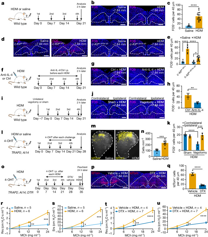
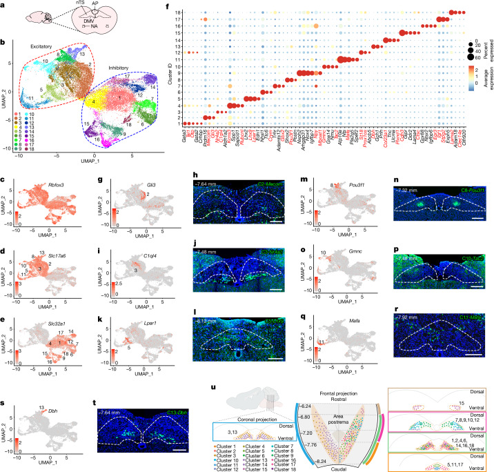
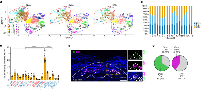
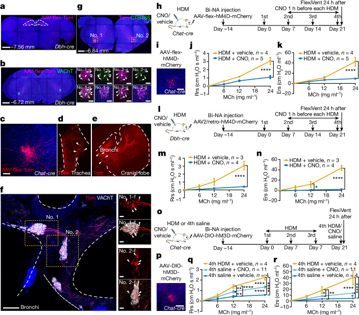
Exaggerated airway constriction triggered by repeated exposure to allergen, also called hyperreactivity, is a hallmark of asthma. Whereas vagal sensory neurons are known to function in allergen-induced hyperreactivity, the identity of downstream nodes remains poorly understood. Here we mapped a full allergen circuit from the lung to the brainstem and back to the lung. Repeated exposure of mice to inhaled allergen activated the nuclei of solitary tract (nTS) neurons in a mast cell-, interleukin-4 (IL-4)- and vagal nerve-dependent manner. Single-nucleus RNA sequencing, followed by RNAscope assay at baseline and allergen challenges, showed that a Dbh nTS population is preferentially activated. Ablation or chemogenetic inactivation of Dbh nTS neurons blunted hyperreactivity whereas chemogenetic activation promoted it. Viral tracing indicated that Dbh nTS neurons project to the nucleus ambiguus (NA) and that NA neurons are necessary and sufficient to relay allergen signals to postganglionic neurons that directly drive airway constriction. Delivery of noradrenaline antagonists to the NA blunted hyperreactivity, suggesting noradrenaline as the transmitter between Dbh nTS and NA. Together, these findings provide molecular, anatomical and functional definitions of key nodes of a canonical allergen response circuit. This knowledge informs how neural modulation could be used to control allergen-induced airway hyperreactivity.
反复接触过敏原引发的气道过度收缩,也称为高反应性,是哮喘的一个标志。虽然已知迷走感觉神经元在过敏原诱导的高反应性中发挥作用,但下游节点的身份仍知之甚少。在这里,我们绘制了一条从肺到脑干再回到肺的完整过敏原通路。将小鼠反复暴露于吸入的过敏原中,以肥大细胞、白细胞介素-4(IL-4)和迷走神经依赖的方式激活孤束核(nTS)神经元的细胞核。单核RNA测序,随后在基线和过敏原刺激下进行RNAscope分析,结果显示Dbh nTS群体被优先激活。Dbh nTS神经元的消融或化学遗传失活减弱了高反应性,而化学遗传激活则增强了高反应性。病毒示踪表明,Dbh nTS神经元投射到疑核(NA),并且NA神经元对于将过敏原信号传递给直接驱动气道收缩的节后神经元是必要且充分的。向NA递送去甲肾上腺素拮抗剂减弱了高反应性,表明去甲肾上腺素是Dbh nTS和NA之间的神经递质。总之,这些发现提供了经典过敏原反应通路关键节点的分子、解剖和功能定义。这些知识为如何利用神经调节来控制过敏原诱导的气道高反应性提供了依据。