Tianjin Medical University Cancer Institute and Hospital, National Clinical Research Center for Cancer, Tianjin, China.
Key Laboratory of Cancer Prevention and Therapy, Tianjin, China.
Cancer Med. 2024 Jul;13(14):e70011. doi: 10.1002/cam4.70011.
Immunotherapy, specifically immune checkpoint inhibitors (ICIs), has revolutionized cancer treatment. However, it can also cause immune-related adverse events (irAEs). This study aimed to develop a clinically practical animal model of irAEs using BALB/c mice.
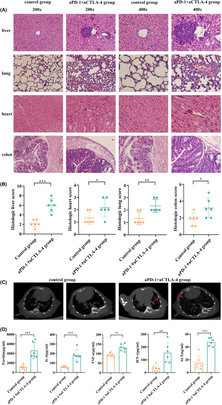
Subcutaneous tumors of mouse breast cancer 4T1 cells were generated in inbred BALB/c mice. The mice were treated with programmed death-1 (PD-1) and cytotoxic t-lymphocyte antigen 4 (CTLA-4) inhibitors once every 3 days for five consecutive administration cycles. Changes in tumor volume and body weight were recorded. Lung computed tomography (CT) scans were conducted. The liver, lungs, heart, and colon tissues of the mice were stained with hematoxylin-eosin (H&E) staining to observe inflammatory infiltration and were scored. Serum samples were collected, and enzyme-linked immunosorbent assay (ELISA) was used to detect the levels of ferritin, glutamic-pyruvic transaminase (ALT), tumor necrosis factor-α (TNF-α), interferon-gamma (IFN-γ), and interleukin-6 (IL-6). Mouse liver and lung cell suspensions were prepared, and changes in macrophages, T cells, myeloid-derived suppressor cells (MDSCs), and regulatory (Treg) cells were detected by flow cytometry.
Mice treated with PD-1 and CTLA-4 inhibitors showed significant reductions in tumor volume and body weight. The tissue inflammatory scores in the experimental group were significantly higher than those in the control group. Lung CT scans of mice in the experimental group showed obvious inflammatory spots. Serum levels of ferritin, IL-6, TNF-α, IFN-γ, and ALT were significantly elevated in the experimental group. Flow cytometry analysis revealed a substantial increase in CD3T cells, Treg cells, and macrophages in the liver and lung tissues of mice in the experimental group compared with the control group, and the change trend of MDSCs was opposite.
The irAE-related animal model was successfully established in BALB/c mice using a combination of PD-1 and CTLA-4 inhibitors through multiple administrations with clinical translational value and practical. This model offers valuable insights into irAE mechanisms for further investigation.
免疫疗法,特别是免疫检查点抑制剂(ICIs),彻底改变了癌症的治疗方式。然而,它也会引起免疫相关不良反应(irAEs)。本研究旨在使用 BALB/c 小鼠建立一种具有临床实用价值的 irAEs 动物模型。
在近交系 BALB/c 小鼠中生成小鼠乳腺癌 4T1 细胞的皮下肿瘤。将小鼠用程序性死亡受体 1(PD-1)和细胞毒性 T 淋巴细胞相关抗原 4(CTLA-4)抑制剂治疗,每 3 天一次,连续 5 个给药周期。记录肿瘤体积和体重的变化。对小鼠进行肺部计算机断层扫描(CT)检查。用苏木精-伊红(H&E)染色对小鼠的肝、肺、心脏和结肠组织进行染色,观察炎症浸润并进行评分。收集血清样本,用酶联免疫吸附测定(ELISA)检测铁蛋白、谷丙转氨酶(ALT)、肿瘤坏死因子-α(TNF-α)、干扰素-γ(IFN-γ)和白细胞介素-6(IL-6)的水平。制备小鼠肝和肺细胞悬液,用流式细胞术检测巨噬细胞、T 细胞、髓源性抑制细胞(MDSCs)和调节性(Treg)细胞的变化。
用 PD-1 和 CTLA-4 抑制剂治疗的小鼠肿瘤体积和体重明显减小。实验组的组织炎症评分明显高于对照组。实验组小鼠的肺部 CT 扫描显示出明显的炎症点。实验组血清铁蛋白、IL-6、TNF-α、IFN-γ和 ALT 水平明显升高。流式细胞术分析显示,实验组小鼠肝、肺组织中 CD3T 细胞、Treg 细胞和巨噬细胞明显增加,而 MDSCs 的变化趋势相反。
通过多次给予 PD-1 和 CTLA-4 抑制剂,成功在 BALB/c 小鼠中建立了具有临床转化价值和实际意义的 irAE 相关动物模型。该模型为进一步研究 irAE 机制提供了有价值的见解。