Jost Tyler A, Gardner Andrea L, Morgan Daylin, Brock Amy
Department of Biomedical Engineering, The University of Texas at Austin.
bioRxiv. 2024 Jul 4:2024.07.02.601576. doi: 10.1101/2024.07.02.601576.
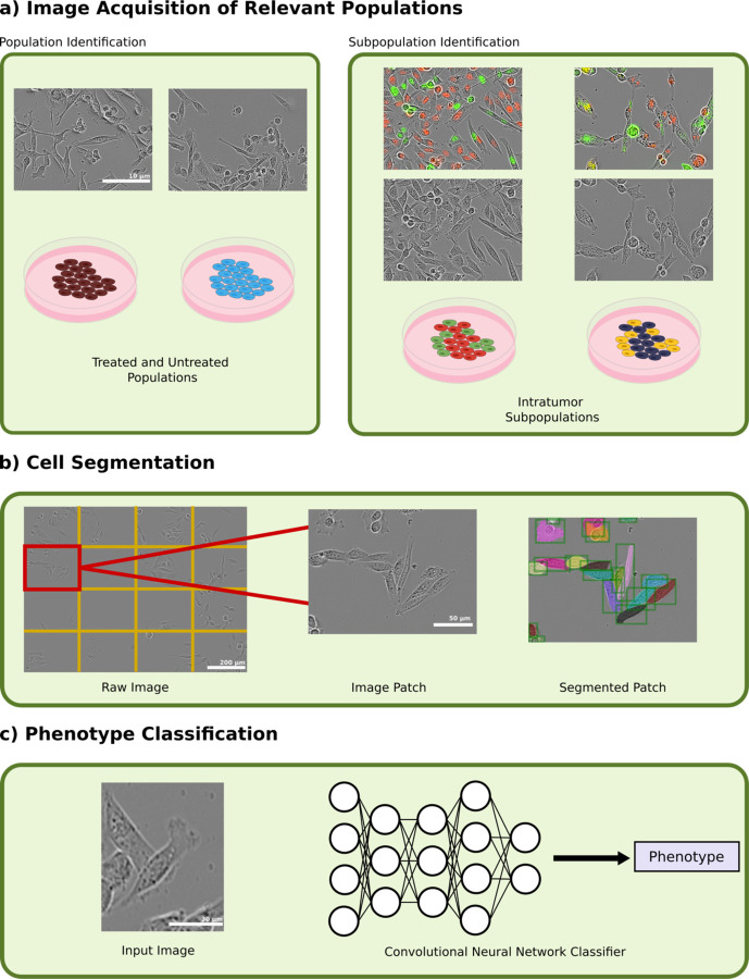
Cells exhibit a wide array of morphological features, enabling computer vision methods to identify and track relevant parameters. Morphological analysis has long been implemented to identify specific cell types and cell responses. Here we asked whether morphological features might also be used to classify transcriptomic subpopulations within cancer cell lines. Identifying cell subpopulations furthers our understanding of morphology as a reflection of underlying cell phenotype and could enable a better understanding of how subsets of cells compete and cooperate in disease progression and treatment.
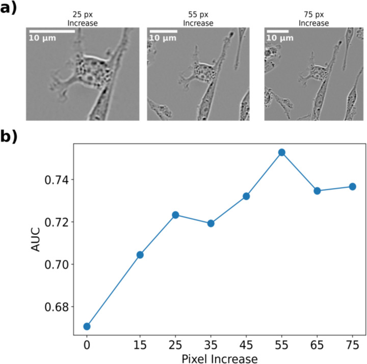
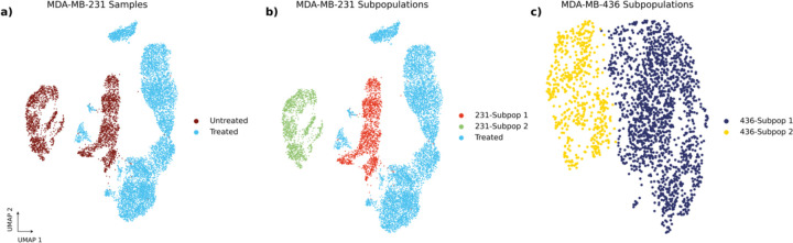
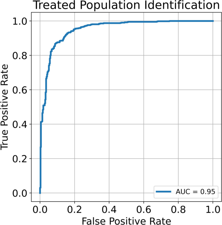
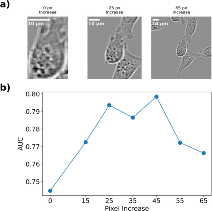
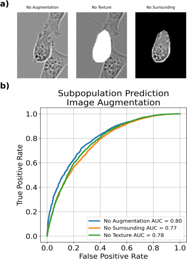
We demonstrate that cell morphology can reflect underlying transcriptomic differences using convolutional neural networks. First, we find that changes induced by chemotherapy treatment are highly identifiable in a breast cancer cell line. We then show that the intra cell line subpopulations that comprise breast cancer cell lines under standard growth conditions are also identifiable using cell morphology. We find that cell morphology is influenced by neighborhood effects beyond the cell boundary, and that including image information surrounding the cell can improve model discrimination ability.
细胞呈现出各种各样的形态特征,这使得计算机视觉方法能够识别和跟踪相关参数。长期以来,形态学分析一直被用于识别特定的细胞类型和细胞反应。在这里,我们探讨形态特征是否也可用于对癌细胞系中的转录组亚群进行分类。识别细胞亚群有助于我们进一步理解形态学是潜在细胞表型的反映,并能够更好地理解细胞亚群在疾病进展和治疗过程中是如何竞争与合作的。
我们证明,使用卷积神经网络,细胞形态能够反映潜在的转录组差异。首先,我们发现在一种乳腺癌细胞系中,化疗诱导的变化是高度可识别的。然后我们表明,在标准生长条件下构成乳腺癌细胞系的细胞系内亚群也可通过细胞形态识别出来。我们发现细胞形态受到细胞边界之外的邻域效应影响,并且纳入细胞周围的图像信息可以提高模型的辨别能力。