• 文献检索
  • 文档翻译
  • 深度研究
  • 学术资讯
  • Suppr Zotero 插件Zotero 插件
  • 邀请有礼
  • 套餐&价格
  • 历史记录
应用&插件
Suppr Zotero 插件Zotero 插件浏览器插件Mac 客户端Windows 客户端微信小程序
定价
高级版会员购买积分包购买API积分包
服务
文献检索文档翻译深度研究API 文档MCP 服务
关于我们
关于 Suppr公司介绍联系我们用户协议隐私条款
关注我们

Suppr 超能文献

核心技术专利:CN118964589B侵权必究
粤ICP备2023148730 号-1Suppr @ 2026

文献检索

告别复杂PubMed语法,用中文像聊天一样搜索,搜遍4000万医学文献。AI智能推荐,让科研检索更轻松。

立即免费搜索

文件翻译

保留排版,准确专业,支持PDF/Word/PPT等文件格式,支持 12+语言互译。

免费翻译文档

深度研究

AI帮你快速写综述,25分钟生成高质量综述,智能提取关键信息,辅助科研写作。

立即免费体验

西尼罗河病毒诱导衰老基因表达调控大脑皮层小胶质细胞表型。

West Nile Virus-Induced Expression of Senescent Gene Regulates Microglial Phenotype within Cerebral Cortex.

机构信息

Center for Neuroimmunology & Neuroinfectious Diseases, St. Louis, MO 63110, USA.

Department of Medicine, Washington University School of Medicine, St. Louis, MO 63110, USA.

出版信息

Biomolecules. 2024 Jul 8;14(7):808. doi: 10.3390/biom14070808.

DOI:10.3390/biom14070808
PMID:39062523
原文链接:https://pmc.ncbi.nlm.nih.gov/articles/PMC11274721/
Abstract

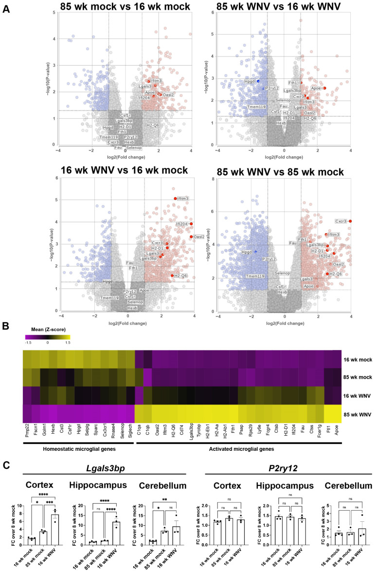
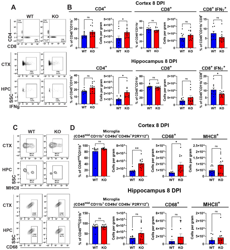
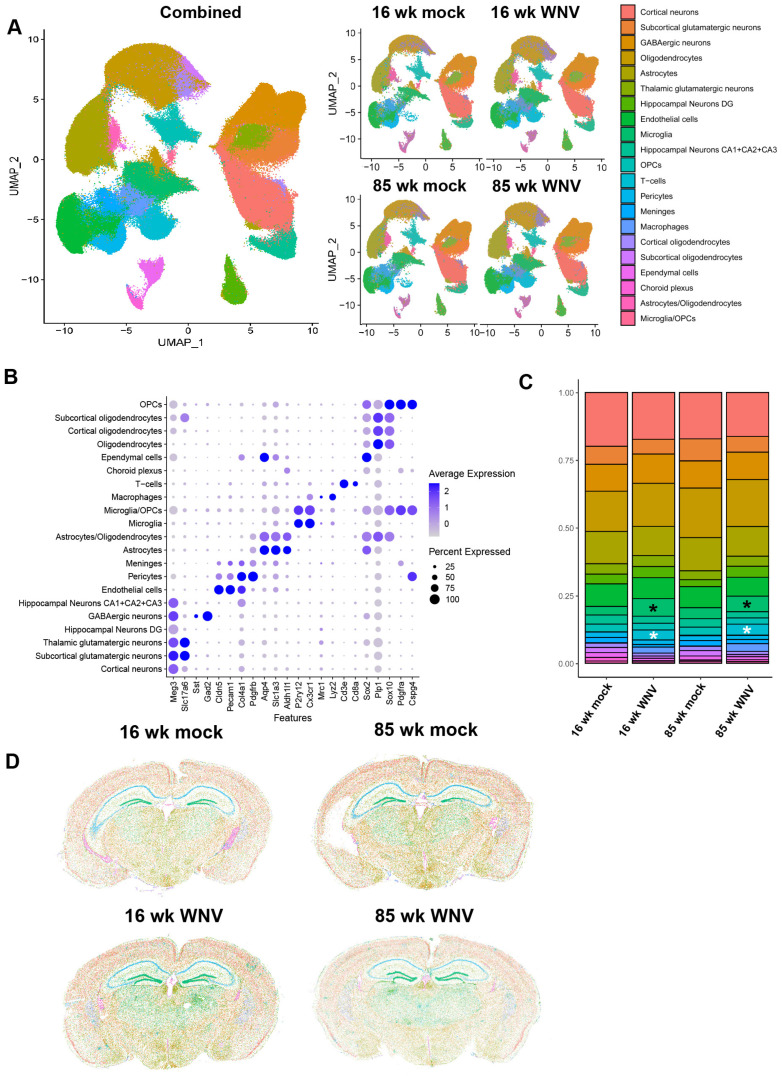
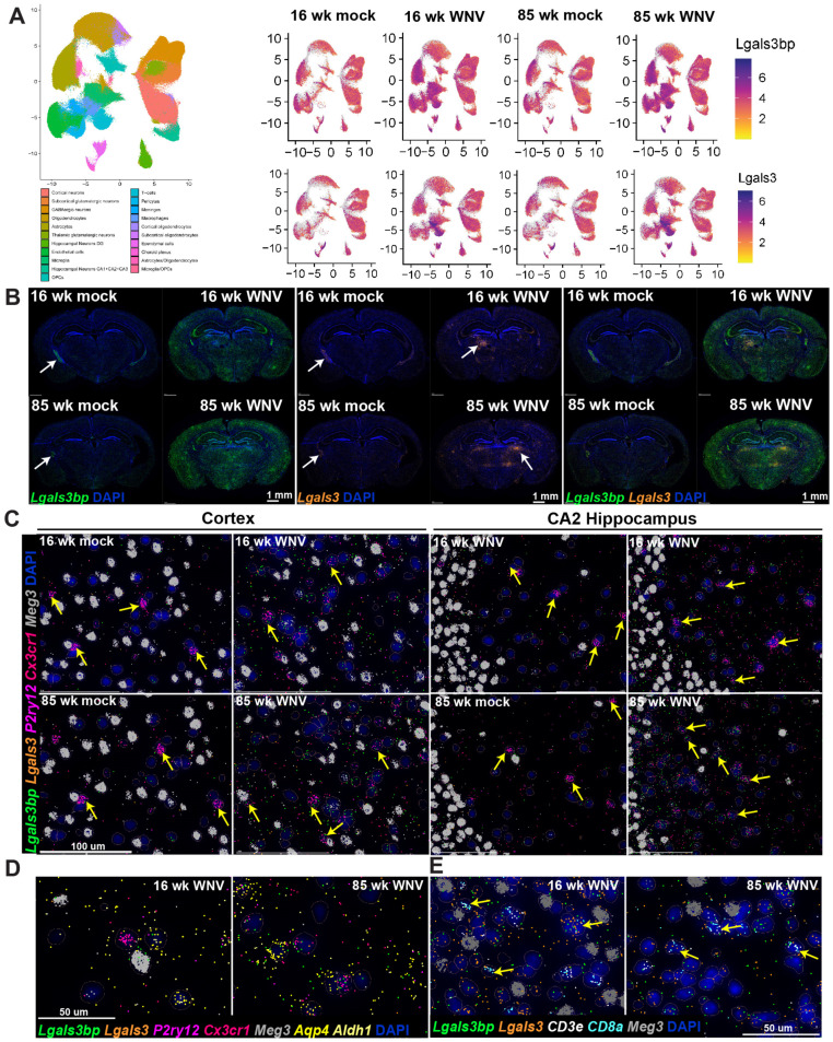
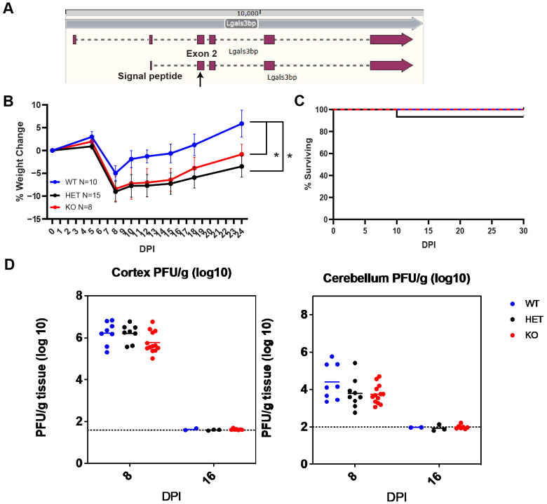
Microglia, the resident macrophages of the central nervous system, exhibit altered gene expression in response to various neurological conditions. This study investigates the relationship between West Nile Virus infection and microglial senescence, focusing on the role of LGALS3BP, a protein implicated in both antiviral responses and aging. Using spatial transcriptomics, RNA sequencing and flow cytometry, we characterized changes in microglial gene signatures in adult and aged mice following recovery from WNV encephalitis. Additionally, we analyzed expression and generated -deficient mice to assess the impact on neuroinflammation and microglial phenotypes. Our results show that WNV-activated microglia share transcriptional signatures with aged microglia, including upregulation of genes involved in interferon response and inflammation. was broadly expressed in the CNS and robustly upregulated during WNV infection and aging. -deficient mice exhibited reduced neuroinflammation, increased homeostatic microglial numbers, and altered T cell populations without differences in virologic control or survival. These data indicate that LGALS3BP has a role in regulating neuroinflammation and microglial activation and suggest that targeting LGALS3BP might provide a potential route for mitigating neuroinflammation-related cognitive decline in aging and post-viral infections.

摘要

小胶质细胞是中枢神经系统的固有巨噬细胞,其基因表达在各种神经疾病中会发生改变。本研究探讨了西尼罗河病毒(WNV)感染与小胶质细胞衰老之间的关系,重点研究了 LGALS3BP 这一在抗病毒反应和衰老中都起作用的蛋白质。本研究使用空间转录组学、RNA 测序和流式细胞术,对成年和老年小鼠在从 WNV 脑炎中恢复后,其小胶质细胞基因特征的变化进行了表征。此外,本研究还分析了 表达情况并生成了 -缺陷小鼠,以评估其对神经炎症和小胶质细胞表型的影响。研究结果表明,WNV 激活的小胶质细胞与衰老的小胶质细胞具有相似的转录特征,包括参与干扰素反应和炎症的基因上调。LGALS3BP 在中枢神经系统中广泛表达,在 WNV 感染和衰老过程中显著上调。LGALS3BP 缺陷小鼠表现出神经炎症减少、稳态小胶质细胞数量增加和 T 细胞群体改变,而病毒学控制或存活方面没有差异。这些数据表明 LGALS3BP 在调节神经炎症和小胶质细胞激活方面具有作用,并提示靶向 LGALS3BP 可能为减轻衰老和病毒性感染相关的认知衰退提供一种潜在途径。

https://cdn.ncbi.nlm.nih.gov/pmc/blobs/4d65/11274721/2db2915e9d73/biomolecules-14-00808-g005.jpg
https://cdn.ncbi.nlm.nih.gov/pmc/blobs/4d65/11274721/bdcdcc257d78/biomolecules-14-00808-g001.jpg
https://cdn.ncbi.nlm.nih.gov/pmc/blobs/4d65/11274721/39358d933fe2/biomolecules-14-00808-g002.jpg
https://cdn.ncbi.nlm.nih.gov/pmc/blobs/4d65/11274721/c2ba8ab3afe3/biomolecules-14-00808-g003.jpg
https://cdn.ncbi.nlm.nih.gov/pmc/blobs/4d65/11274721/b0d1a731fab9/biomolecules-14-00808-g004.jpg
https://cdn.ncbi.nlm.nih.gov/pmc/blobs/4d65/11274721/2db2915e9d73/biomolecules-14-00808-g005.jpg

相似文献

1
West Nile Virus-Induced Expression of Senescent Gene Regulates Microglial Phenotype within Cerebral Cortex.西尼罗河病毒诱导衰老基因表达调控大脑皮层小胶质细胞表型。
Biomolecules. 2024 Jul 8;14(7):808. doi: 10.3390/biom14070808.
2
Minocycline Has Anti-inflammatory Effects and Reduces Cytotoxicity in an Spinal Cord Slice Culture Model of West Nile Virus Infection.米诺环素在西尼罗河病毒感染的脊髓切片培养模型中具有抗炎作用并降低细胞毒性。
J Virol. 2017 Oct 27;91(22). doi: 10.1128/JVI.00569-17. Print 2017 Nov 15.
3
Peli1 facilitates virus replication and promotes neuroinflammation during West Nile virus infection.Peli1 促进西尼罗河病毒感染期间的病毒复制和神经炎症。
J Clin Invest. 2018 Nov 1;128(11):4980-4991. doi: 10.1172/JCI99902. Epub 2018 Sep 24.
4
CSF1R antagonism limits local restimulation of antiviral CD8 T cells during viral encephalitis.CSF1R 拮抗作用限制了病毒性脑炎期间抗病毒 CD8 T 细胞的局部再刺激。
J Neuroinflammation. 2019 Jan 31;16(1):22. doi: 10.1186/s12974-019-1397-4.
5
Treatment with Granulocyte-Macrophage Colony-Stimulating Factor Reduces Viral Titers in the Brains of West Nile Virus-Infected Mice and Improves Survival.粒细胞-巨噬细胞集落刺激因子治疗可降低西尼罗河病毒感染小鼠脑中的病毒滴度并提高存活率。
J Virol. 2023 Mar 30;97(3):e0180522. doi: 10.1128/jvi.01805-22. Epub 2023 Feb 21.
6
MAVS Expressed by Hematopoietic Cells Is Critical for Control of West Nile Virus Infection and Pathogenesis.造血细胞表达的MAVS对控制西尼罗河病毒感染和发病机制至关重要。
J Virol. 2016 Jul 27;90(16):7098-7108. doi: 10.1128/JVI.00707-16. Print 2016 Aug 15.
7
The Interferon-Stimulated Gene Ifi27l2a Restricts West Nile Virus Infection and Pathogenesis in a Cell-Type- and Region-Specific Manner.干扰素刺激基因Ifi27l2a以细胞类型和区域特异性方式限制西尼罗河病毒感染和发病机制。
J Virol. 2015 Dec 23;90(5):2600-15. doi: 10.1128/JVI.02463-15.
8
Intrinsic Innate Immune Responses Control Viral Growth and Protect against Neuronal Death in an Model of West Nile Virus-Induced Central Nervous System Disease.内在先天免疫反应控制病毒生长并防止西尼罗河病毒诱导的中枢神经系统疾病模型中的神经元死亡。
J Virol. 2021 Aug 25;95(18):e0083521. doi: 10.1128/JVI.00835-21.
9
Cellular microRNA-155 Regulates Virus-Induced Inflammatory Response and Protects against Lethal West Nile Virus Infection.细胞微小RNA-155调控病毒诱导的炎症反应并抵御西尼罗河病毒致死性感染。
Viruses. 2019 Dec 19;12(1):9. doi: 10.3390/v12010009.
10
CCR5 limits cortical viral loads during West Nile virus infection of the central nervous system.在西尼罗河病毒感染中枢神经系统期间,CCR5限制皮质病毒载量。
J Neuroinflammation. 2015 Dec 15;12:233. doi: 10.1186/s12974-015-0447-9.

引用本文的文献

1
CXCR4 and CXCR6 dually limit T cell entry into the polyomavirus-infected brain.CXCR4和CXCR6双重限制T细胞进入多瘤病毒感染的大脑。
J Neuroinflammation. 2025 Jun 28;22(1):169. doi: 10.1186/s12974-025-03496-2.
2
Active Cryptococcus neoformans glucuronoxylomannan production prevents elimination of cryptococcal CNS infection in vivo.新型隐球菌活性葡糖醛酸木聚糖甘露聚糖的产生可阻止体内隐球菌中枢神经系统感染的清除。
J Neuroinflammation. 2025 Mar 4;22(1):61. doi: 10.1186/s12974-025-03384-9.
3
Protein kinase R induced by type I interferons is a main regulator of reactive microglia in Zika virus infection.

本文引用的文献

1
Microglial Senescence and Activation in Healthy Aging and Alzheimer's Disease: Systematic Review and Neuropathological Scoring.小胶质细胞衰老和激活在健康衰老和阿尔茨海默病中的作用:系统评价和神经病理学评分。
Cells. 2023 Dec 12;12(24):2824. doi: 10.3390/cells12242824.
2
Molecular Mechanisms Associated with Neurodegeneration of Neurotropic Viral Infection.与嗜神经病毒感染所致神经退行性变相关的分子机制
Mol Neurobiol. 2024 May;61(5):2881-2903. doi: 10.1007/s12035-023-03761-6. Epub 2023 Nov 9.
3
Transcriptional and epigenetic decoding of the microglial aging process.
I型干扰素诱导的蛋白激酶R是寨卡病毒感染中反应性小胶质细胞的主要调节因子。
Glia. 2025 Jan;73(1):80-104. doi: 10.1002/glia.24619. Epub 2024 Oct 3.
解析小胶质细胞衰老过程中的转录和表观遗传。
Nat Aging. 2023 Oct;3(10):1288-1311. doi: 10.1038/s43587-023-00479-x. Epub 2023 Sep 11.
4
Extracellular LGALS3BP: a potential disease marker and actionable target for antibody-drug conjugate therapy in glioblastoma.细胞外 LGALS3BP:胶质母细胞瘤中抗体药物偶联物治疗的潜在疾病标志物和作用靶点。
Mol Oncol. 2023 Aug;17(8):1460-1473. doi: 10.1002/1878-0261.13453. Epub 2023 Jun 7.
5
Aging microglia.衰老的小胶质细胞。
Cell Mol Life Sci. 2023 Apr 21;80(5):126. doi: 10.1007/s00018-023-04775-y.
6
Microglia at the scene of the crime: what their transcriptomics reveal about brain health.小胶质细胞在犯罪现场:它们的转录组学如何揭示大脑健康。
Curr Opin Neurol. 2023 Jun 1;36(3):207-213. doi: 10.1097/WCO.0000000000001151. Epub 2023 Apr 18.
7
Viral pathogens increase risk of neurodegenerative disease.病毒病原体增加神经退行性疾病的风险。
Nat Rev Neurol. 2023 May;19(5):259-260. doi: 10.1038/s41582-023-00790-6.
8
Molecular and spatial signatures of mouse brain aging at single-cell resolution.单细胞分辨率解析的小鼠大脑衰老的分子和空间特征。
Cell. 2023 Jan 5;186(1):194-208.e18. doi: 10.1016/j.cell.2022.12.010. Epub 2022 Dec 28.
9
Single-cell RNA transcriptome analysis of CNS immune cells reveals CXCL16/CXCR6 as maintenance factors for tissue-resident T cells that drive synapse elimination.中枢神经系统免疫细胞的单细胞转录组分析显示,CXCL16/CXCR6 可作为组织驻留 T 细胞的维持因子,促进突触消除。
Genome Med. 2022 Sep 24;14(1):108. doi: 10.1186/s13073-022-01111-0.
10
A new gene set identifies senescent cells and predicts senescence-associated pathways across tissues.一组新的基因集可识别衰老细胞,并预测跨组织的衰老相关途径。
Nat Commun. 2022 Aug 16;13(1):4827. doi: 10.1038/s41467-022-32552-1.