Vijayakumar Periyasamy, Mishra Anamika, Deka Ram Pratim, Pinto Sneha M, Subbannayya Yashwanth, Sood Richa, Prasad Thottethodi Subrahmanya Keshava, Raut Ashwin Ashok
Pathogenomics Laboratory, WOAH Reference Lab for Avian Influenza, ICAR-National Institute of High Security Animal Diseases, Bhopal 462022, Madhya Pradesh, India.
Veterinary College and Research Institute, Tamil Nadu Veterinary and Animal Sciences University, Salem 600051, Tamil Nadu, India.
Microorganisms. 2024 Jun 25;12(7):1288. doi: 10.3390/microorganisms12071288.
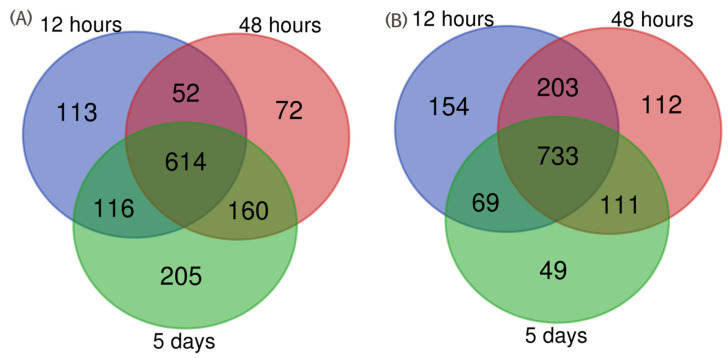
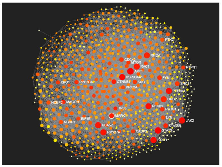
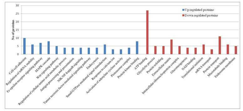
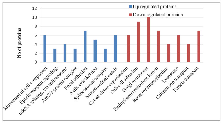
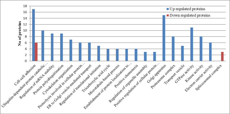
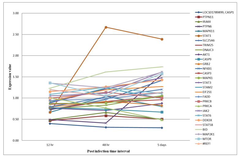
Domestic ducks () are resistant to most of the highly pathogenic avian influenza virus (HPAIV) infections. In this study, we characterized the lung proteome and phosphoproteome of ducks infected with the HPAI H5N1 virus (A/duck/India/02CA10/2011/Agartala) at 12 h, 48 h, and 5 days post-infection. A total of 2082 proteins were differentially expressed and 320 phosphorylation sites mapping to 199 phosphopeptides, corresponding to 129 proteins were identified. The functional annotation of the proteome data analysis revealed the activation of the RIG-I-like receptor and Jak-STAT signaling pathways, which led to the induction of interferon-stimulated gene (ISG) expression. The pathway analysis of the phosphoproteome datasets also confirmed the activation of RIG-I, Jak-STAT signaling, NF-kappa B signaling, and MAPK signaling pathways in the lung tissues. The induction of ISG proteins (STAT1, STAT3, STAT5B, STAT6, IFIT5, and PKR) established a protective anti-viral immune response in duck lung tissue. Further, the protein-protein interaction network analysis identified proteins like AKT1, STAT3, JAK2, RAC1, STAT1, PTPN11, RPS27A, NFKB1, and MAPK1 as the main hub proteins that might play important roles in disease progression in ducks. Together, the functional annotation of the proteome and phosphoproteome datasets revealed the molecular basis of the disease progression and disease resistance mechanism in ducks infected with the HPAI H5N1 virus.
家鸭对大多数高致病性禽流感病毒(HPAIV)感染具有抵抗力。在本研究中,我们对感染HPAI H5N1病毒(A/duck/India/02CA10/2011/Agartala)12小时、48小时和感染后5天的鸭肺蛋白质组和磷酸化蛋白质组进行了表征。共鉴定出2082种差异表达蛋白以及320个磷酸化位点,这些位点映射到199个磷酸肽,对应129种蛋白质。蛋白质组数据分析的功能注释显示,维甲酸诱导基因I样受体和Jak-STAT信号通路被激活,这导致了干扰素刺激基因(ISG)表达的诱导。磷酸化蛋白质组数据集的通路分析也证实了肺组织中维甲酸诱导基因I、Jak-STAT信号、核因子κB信号和丝裂原活化蛋白激酶信号通路的激活。ISG蛋白(STAT1、STAT3、STAT5B、STAT6、IFIT5和PKR)的诱导在鸭肺组织中建立了保护性抗病毒免疫反应。此外,蛋白质-蛋白质相互作用网络分析确定了AKT1、STAT3、JAK2、RAC1、STAT1、PTPN11、RPS27A、NFKB1和MAPK1等蛋白质为主要枢纽蛋白,它们可能在鸭疾病进展中起重要作用。总之,蛋白质组和磷酸化蛋白质组数据集的功能注释揭示了感染HPAI H5N1病毒的鸭疾病进展和抗病机制的分子基础。