Shalaby Asem, Shalaby Ola, Abdullah Hazem, Boulassel Mohamed Rachid, Arafa Mohammad
Pathology Department, Faculty of Medicine,, Mansoura University, Mansoura, Egypt.
Pathology Department, College of Medicine and Health Sciences, Sultan Qaboos University, Muscat, Oman.
Clin Transl Oncol. 2025 Feb;27(2):699-705. doi: 10.1007/s12094-024-03613-2. Epub 2024 Aug 1.
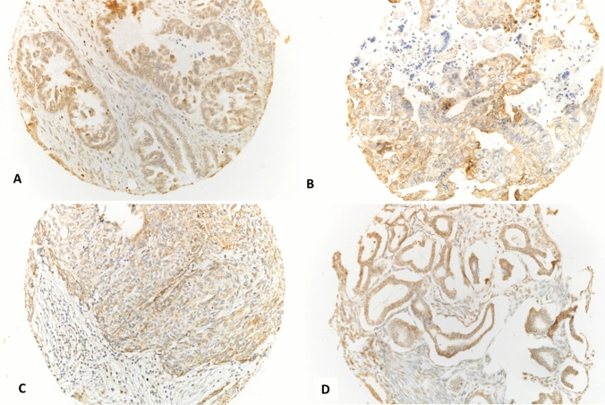
Primary carcinoma of the ovary (OCs) are responsible for a significant number of deaths related to cancer, and have the highest rate of death related to cancers of the female reproductive organs. Programmed cell death 1 (PD1) protein, acts as an immune checkpoint, and has an important role in the down-regulation of the immune system by preventing the activation of T-cells, which will weaken the autoimmunity and increases self-tolerance. This study aimed at the evaluation of the immunohistochemical (IHC) expression of PD-L1 in various primary surface ovarian epithelial tumours and to test its correlation with different clinicopathological parameters together with the expression of a panel of P53, ER and PR.
A set of 102 cases of primary ovarian surface epithelial neoplasms (benign, borderline and malignant) were collected to construct Tissue Microarray (TMA) using 3 tissue cores from each case. IHC for PD-L1, p53, PR and ER was performed. The expression of PD-L1 was evaluated in relation to some clinicopathological parameters and to the expression patterns of other markers.
Expression of PD-L1 was detected in about 51% (n = 36) of malignant tumours. The malignant group significantly showed PD-L1 positivity compared to borderline and benign groups. The malignant tumours significantly showed PD-L1 and total p53 positivity in comparison to borderline group. Also, malignant tumours significantly showed higher combined positivity of PD-L1 and either PR or ER compared to borderline and benign lesions. No significant correlation was appreciated between PD-L1 expression and with any of the studied clinicopathological parameters.
This study showed a significant PD-L1 expression in malignant primary surface epithelial tumours. Construction of a panel of IHC markers, including PD-L1, could have a potential value to define patients those would benefit from the addition of immunotherapy to the treatment plan.
原发性卵巢癌(OCs)导致了大量癌症相关死亡,在女性生殖器官癌症中死亡率最高。程序性细胞死亡蛋白1(PD1)作为一种免疫检查点,通过阻止T细胞激活在免疫系统下调中起重要作用,这会削弱自身免疫并增加自我耐受性。本研究旨在评估程序性死亡受体配体1(PD-L1)在各种原发性卵巢表面上皮肿瘤中的免疫组化(IHC)表达,并测试其与不同临床病理参数以及一组P53、雌激素受体(ER)和孕激素受体(PR)表达的相关性。
收集102例原发性卵巢表面上皮肿瘤(良性、交界性和恶性)病例,每例取3个组织芯构建组织芯片(TMA)。进行PD-L1、p53、PR和ER的免疫组化检测。评估PD-L1的表达与一些临床病理参数以及其他标志物表达模式的关系。
在约51%(n = 36)的恶性肿瘤中检测到PD-L1表达。与交界性和良性组相比,恶性组显著显示PD-L1阳性。与交界性组相比,恶性肿瘤显著显示PD-L1和总p53阳性。此外,与交界性和良性病变相比,恶性肿瘤显著显示PD-L1与PR或ER的联合阳性更高。未发现PD-L1表达与任何研究的临床病理参数之间存在显著相关性。
本研究显示恶性原发性表面上皮肿瘤中PD-L1表达显著。构建包括PD-L1在内的免疫组化标志物组合可能对确定那些将从治疗方案中添加免疫治疗中受益的患者具有潜在价值。