• 文献检索
  • 文档翻译
  • 深度研究
  • 学术资讯
  • Suppr Zotero 插件Zotero 插件
  • 邀请有礼
  • 套餐&价格
  • 历史记录
应用&插件
Suppr Zotero 插件Zotero 插件浏览器插件Mac 客户端Windows 客户端微信小程序
定价
高级版会员购买积分包购买API积分包
服务
文献检索文档翻译深度研究API 文档MCP 服务
关于我们
关于 Suppr公司介绍联系我们用户协议隐私条款
关注我们

Suppr 超能文献

核心技术专利:CN118964589B侵权必究
粤ICP备2023148730 号-1Suppr @ 2026

文献检索

告别复杂PubMed语法,用中文像聊天一样搜索,搜遍4000万医学文献。AI智能推荐,让科研检索更轻松。

立即免费搜索

文件翻译

保留排版,准确专业,支持PDF/Word/PPT等文件格式,支持 12+语言互译。

免费翻译文档

深度研究

AI帮你快速写综述,25分钟生成高质量综述,智能提取关键信息,辅助科研写作。

立即免费体验

上皮性卵巢肿瘤:临床病理相关性及免疫组化研究

Epithelial ovarian tumors: Clinicopathological correlation and immunohistochemical study.

作者信息

Naik Pooja S, Deshmukh Sanjay, Khandeparkar Siddhi Gaurish Sinai, Joshi Avinash, Babanagare Shridhar, Potdar Jyostna, Risbud Neelesh Sharad

机构信息

Department of Pathology, Shrimati Kashibai Navale Medical College and General Hospital, Pune, Maharashtra, India.

Department of Obstetrics and Gynaecology, Shrimati Kashibai Navale Medical College and General Hospital, Pune, Maharashtra, India.

出版信息

J Midlife Health. 2015 Oct-Dec;6(4):178-83. doi: 10.4103/0976-7800.172349.

DOI:10.4103/0976-7800.172349
PMID:26903759
原文链接:https://pmc.ncbi.nlm.nih.gov/articles/PMC4743281/
Abstract

BACKGROUND

Ovarian cancer is the third leading site of cancer among women, trailing behind cervix and breast cancer.

AIM

This study was undertaken to analyze the immunohistochemical (IHC) profile of estrogen receptors (ER), progesterone receptors (PR), Ki-67, and p53 in various ovarian epithelial tumors and attempt correlation with clinical and histopathological findings.

MATERIALS AND METHODS

The present study was conducted over a period of 4 years. A technique of manual tissue array was employed for cases subjected for IHC. The primary antibodies used were ER, PR, p53, and Ki-67. A correlation was attempted between histopathological and IHC findings. Results were subjected to statistical analysis. Software program "the primer of biostatistics 5.0" was used for calculation of interrelationships between the analyzed ER, PR, p53, and Ki-67 expression and histological factors by Pearson's Chi-square test. The results were considered to be significant when the P < 0.05.

RESULTS

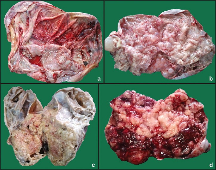
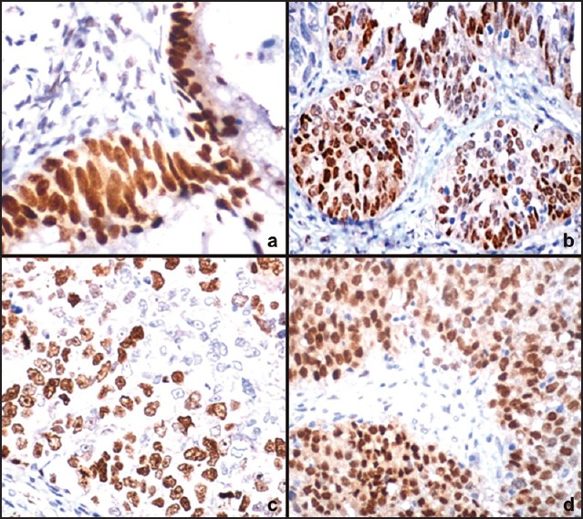
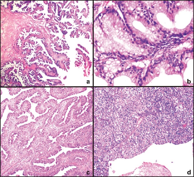
There were 110 cases of surface epithelial ovarian tumors (SEOT) encountered over the period of 4 years. The expression of ER was more in malignant tumors (13/16, 81.25%) than borderline (9/12, 75%) and benign (20/82, 24.39%). As compared to ER, the expression of PR was more in benign (51/82, 62.19%) than borderline (8/12, 66.67%) and malignant tumors (9/16, 56.25%). The expression of PR was more in benign tumors than borderline and malignant tumors. However, this was not statistically significant (Chi-square = 0.335 with 2 degrees of freedom; P = 0.846). The expression of p53 was less in benign (5/82, 6.1%) than borderline (9/12, 75%) and malignant tumors (13/16, 81.25%). The expression of Ki-67 was more in malignant (4/82, 4.88%) than borderline (10/12, 83.33%) and benign tumors (15/16, 93.75%). In all the above cases, the difference was statistically significant (P < 0.05). There was statistically significant difference in the expression of ER, PR, p53, and Ki-67 in the patients with age <40 years and above 40 years (P = 0.912). A positive correlation was observed in p53 expression and tumor grade. Similar correlation was seen in Ki-67 and tumor grade. It was also noted that mean Ki-67 labeling index (Li) had also increased with tumor grade. In the case of serous tumors, ER was expressed in all high- and low-grade tumors. The expression of PR was more in low-grade tumors than high-grade ones. P53 expression was seen in all high-grade tumors and 33.34% of low-grade tumor. The Ki-67 Li was more in high-grade tumors than low-grade tumors. Expression of ER, p53, and Ki-67 was higher in tumor showing metastasis. The mean Ki-67 Li was also higher in metastasizing tumors. However, PR expression was less in metastasizing tumors than nonmetastasizing tumors.

CONCLUSION

IHC marker report of ER, PR status, and Ki-67 if included in each pathology report will pave the way for better understanding of biological behavior and modify treatment strategies.

摘要

背景

卵巢癌是女性中第三大常见癌症部位,仅次于宫颈癌和乳腺癌。

目的

本研究旨在分析各种卵巢上皮性肿瘤中雌激素受体(ER)、孕激素受体(PR)、Ki-67和p53的免疫组化(IHC)特征,并尝试将其与临床和组织病理学结果相关联。

材料与方法

本研究历时4年。对进行免疫组化的病例采用手工组织芯片技术。使用的一抗为ER、PR、p53和Ki-67。尝试将组织病理学和免疫组化结果进行关联。结果进行统计学分析。使用软件程序“生物统计学基础5.0”通过Pearson卡方检验计算分析的ER、PR、p53和Ki-67表达与组织学因素之间的相互关系。当P<0.05时,结果被认为具有统计学意义。

结果

在4年期间共遇到110例表面上皮性卵巢肿瘤(SEOT)。ER在恶性肿瘤中的表达(13/16,81.25%)高于交界性肿瘤(9/12,75%)和良性肿瘤(20/82,24.39%)。与ER相比,PR在良性肿瘤中的表达(51/82,62.19%)高于交界性肿瘤(8/12,66.67%)和恶性肿瘤(9/16,56.25%)。PR在良性肿瘤中的表达高于交界性和恶性肿瘤。然而,这在统计学上无显著差异(卡方=0.335,自由度为2;P=0.846)。p53在良性肿瘤中的表达(5/82,6.1%)低于交界性肿瘤(9/12,75%)和恶性肿瘤(13/16,81.25%)。Ki-67在恶性肿瘤中的表达(4/82,4.88%)低于交界性肿瘤(10/12,83.33%)和良性肿瘤(15/16,93.75%)。在上述所有病例中,差异具有统计学意义(P<0.05)。年龄<40岁和>40岁患者的ER、PR、p53和Ki-67表达存在统计学显著差异(P=0.912)。p53表达与肿瘤分级呈正相关。Ki-67与肿瘤分级也有类似的相关性。还注意到平均Ki-67标记指数(Li)也随肿瘤分级增加。在浆液性肿瘤中,ER在所有高分级和低分级肿瘤中均有表达。PR在低分级肿瘤中的表达高于高分级肿瘤。P53表达见于所有高分级肿瘤和33.34%的低分级肿瘤。Ki-67 Li在高分级肿瘤中高于低分级肿瘤。ER、p53和Ki-67在有转移的肿瘤中表达较高。转移肿瘤的平均Ki-67 Li也较高。然而,PR在转移肿瘤中的表达低于非转移肿瘤。

结论

如果在每份病理报告中纳入ER、PR状态和Ki-67的免疫组化标记报告,将为更好地理解生物学行为和调整治疗策略铺平道路。

https://cdn.ncbi.nlm.nih.gov/pmc/blobs/b419/4743281/5a9042b93a3e/JMH-6-178-g003.jpg
https://cdn.ncbi.nlm.nih.gov/pmc/blobs/b419/4743281/aefa812fe9a9/JMH-6-178-g001.jpg
https://cdn.ncbi.nlm.nih.gov/pmc/blobs/b419/4743281/a94b7cc8c41c/JMH-6-178-g002.jpg
https://cdn.ncbi.nlm.nih.gov/pmc/blobs/b419/4743281/5a9042b93a3e/JMH-6-178-g003.jpg
https://cdn.ncbi.nlm.nih.gov/pmc/blobs/b419/4743281/aefa812fe9a9/JMH-6-178-g001.jpg
https://cdn.ncbi.nlm.nih.gov/pmc/blobs/b419/4743281/a94b7cc8c41c/JMH-6-178-g002.jpg
https://cdn.ncbi.nlm.nih.gov/pmc/blobs/b419/4743281/5a9042b93a3e/JMH-6-178-g003.jpg

相似文献

1
Epithelial ovarian tumors: Clinicopathological correlation and immunohistochemical study.上皮性卵巢肿瘤:临床病理相关性及免疫组化研究
J Midlife Health. 2015 Oct-Dec;6(4):178-83. doi: 10.4103/0976-7800.172349.
2
The expression of immunohistochemical markers estrogen receptor, progesterone receptor, Her-2-neu, p53 and Ki-67 in epithelial ovarian tumors and its correlation with clinicopathologic variables.上皮性卵巢肿瘤中雌激素受体、孕激素受体、Her-2-neu、p53和Ki-67免疫组化标志物的表达及其与临床病理变量的相关性。
Indian J Pathol Microbiol. 2012 Jan-Mar;55(1):33-7. doi: 10.4103/0377-4929.94852.
3
Immunohistochemical expression of Ki-67, p53, and CD10 in phyllodes tumor and their correlation with its histological grade.叶状肿瘤中Ki-67、p53和CD10的免疫组化表达及其与组织学分级的相关性。
J Lab Physicians. 2019 Oct-Dec;11(4):330-334. doi: 10.4103/JLP.JLP_106_19.
4
P53, bcl-2, ki-67 li (labeling index) status in benign, proliferative, and malignant ovarian surface epithelial neoplasms.P53、bcl-2、ki-67 标记指数(LI)在良性、增殖性和恶性卵巢表面上皮性肿瘤中的状态。
Eurasian J Med. 2009 Apr;41(1):10-4.
5
The immunohistochemical expression of p53 and Ki67 in ovarian epithelial borderline tumors. Correlation with clinicopathological factors.p53和Ki67在卵巢上皮性交界性肿瘤中的免疫组化表达。与临床病理因素的相关性。
Rom J Morphol Embryol. 2012;53(4):967-73.
6
[Relationships between the molecular biomarkers and the clinicopathologic features and prognosis in endometrial carcinoma].子宫内膜癌中分子生物标志物与临床病理特征及预后的关系
Beijing Da Xue Xue Bao Yi Xue Ban. 2011 Oct 18;43(5):743-8.
7
Study of Immunohistochemical Markers (CK-19, CD-56, Ki-67, p53) in Differentiating Benign and Malignant Solitary Thyroid Nodules with special Reference to Papillary Thyroid Carcinomas.免疫组织化学标志物(CK-19、CD-56、Ki-67、p53)在鉴别甲状腺良恶性孤立性结节中的研究——以甲状腺乳头状癌为重点
J Clin Diagn Res. 2016 Dec;10(12):EC14-EC19. doi: 10.7860/JCDR/2016/22428.9114. Epub 2016 Dec 1.
8
Joint detection of multiple immunohistochemical indices and clinical significance in breast cancer.乳腺癌中多种免疫组化指标的联合检测及临床意义
Mol Clin Oncol. 2013 Jul;1(4):703-710. doi: 10.3892/mco.2013.111. Epub 2013 May 8.
9
Prognostic significance of ER, PR, Ki67, c-erbB-2, and p53 in endometrial carcinoma.雌激素受体、孕激素受体、Ki67、c-erbB-2及p53在子宫内膜癌中的预后意义
J Med Assoc Thai. 2008 Dec;91(12):1779-84.
10
Steroid receptors and hormones in relation to cell proliferation and apoptosis in poorly differentiated epithelial ovarian tumors.类固醇受体和激素与低分化上皮性卵巢肿瘤细胞增殖及凋亡的关系
Int J Oncol. 2001 Jul;19(1):31-8.

引用本文的文献

1
Borderline ovarian tumor in the pediatric and adolescent population: a clinopathologic analysis of fourteen cases.儿童及青少年人群中的卵巢交界性肿瘤:14例临床病理分析
Int J Clin Exp Pathol. 2020 May 1;13(5):1053-1059. eCollection 2020.
2
Different TP53 mutants in p53 overexpressed epithelial ovarian carcinoma can be associated both with altered and unaltered glycolytic and apoptotic profiles.p53过表达的上皮性卵巢癌中的不同TP53突变体可能与糖酵解和凋亡谱的改变及未改变均相关。
Cancer Cell Int. 2018 Jan 30;18:14. doi: 10.1186/s12935-018-0514-2. eCollection 2018.
3
The positivity of estrogen receptor and progesterone receptor may not be associated with metastasis and recurrence in epithelial ovarian cancer.

本文引用的文献

1
Correlation Between Immunohistochemical Biomarkers Expression and Prognosis of Ovarian Carcinomas in Tunisian Patients.突尼斯患者卵巢癌免疫组化生物标志物表达与预后的相关性
World J Oncol. 2010 Jun;1(3):118-128. doi: 10.4021/wjon2010.06.213w. Epub 2010 May 19.
2
P53, bcl-2, ki-67 li (labeling index) status in benign, proliferative, and malignant ovarian surface epithelial neoplasms.P53、bcl-2、ki-67 标记指数(LI)在良性、增殖性和恶性卵巢表面上皮性肿瘤中的状态。
Eurasian J Med. 2009 Apr;41(1):10-4.
3
The expression of immunohistochemical markers estrogen receptor, progesterone receptor, Her-2-neu, p53 and Ki-67 in epithelial ovarian tumors and its correlation with clinicopathologic variables.
雌激素受体和孕激素受体的阳性表达可能与上皮性卵巢癌的转移和复发无关。
Sci Rep. 2017 Dec 5;7(1):16922. doi: 10.1038/s41598-017-17265-6.
4
Hormone receptors expression in ovarian cancer taking into account menopausal status: a retrospective study in Chinese population.考虑绝经状态的卵巢癌中激素受体表达:中国人群的一项回顾性研究
Oncotarget. 2017 Aug 14;8(48):84019-84027. doi: 10.18632/oncotarget.20251. eCollection 2017 Oct 13.
5
MZF-1/Elk-1/PKCα is Associated with Poor Prognosis in Patients with Hepatocellular Carcinoma.MZF-1/Elk-1/PKCα与肝细胞癌患者的不良预后相关。
J Cancer. 2017 Sep 2;8(15):3028-3036. doi: 10.7150/jca.20467. eCollection 2017.
6
The Putative Role of Alterations and p53 Expression in Borderline Ovarian Tumors - Correlation with Clinicopathological Features and Prognosis: A Mini-Review.改变和p53表达在卵巢交界性肿瘤中的假定作用——与临床病理特征及预后的相关性:一篇综述
J Cancer. 2017 Aug 22;8(14):2684-2691. doi: 10.7150/jca.19691. eCollection 2017.
上皮性卵巢肿瘤中雌激素受体、孕激素受体、Her-2-neu、p53和Ki-67免疫组化标志物的表达及其与临床病理变量的相关性。
Indian J Pathol Microbiol. 2012 Jan-Mar;55(1):33-7. doi: 10.4103/0377-4929.94852.
4
Estrogen and progesterone receptor status and outcome in epithelial ovarian cancers and low malignant potential tumors.上皮性卵巢癌及低恶性潜能肿瘤中的雌激素和孕激素受体状态与预后
Gynecol Oncol. 2009 Sep;114(3):480-5. doi: 10.1016/j.ygyno.2009.05.045. Epub 2009 Jun 27.
5
Expression of the estrogen and progesterone receptors as prognostic factor in serous ovarian cancers.雌激素和孕激素受体表达作为浆液性卵巢癌的预后因素
Exp Oncol. 2009 Mar;31(1):48-51.
6
Study of 'patterns of care' of ovarian cancer patients in a specialized cancer institute in Kolkata, eastern India.印度东部加尔各答一家专业癌症研究所中卵巢癌患者的“护理模式”研究。
Indian J Cancer. 2009 Jan-Mar;46(1):28-33. doi: 10.4103/0019-509x.48592.
7
Ovarian carcinogenesis: an alternative hypothesis.卵巢癌发生:一种替代假说。
Adv Exp Med Biol. 2008;622:79-87. doi: 10.1007/978-0-387-68969-2_7.
8
Histologic subtypes of ovarian carcinoma: an overview.卵巢癌的组织学亚型:概述
Int J Gynecol Pathol. 2008 Apr;27(2):161-74. doi: 10.1097/PGP.0b013e31815ea812.
9
Androgen receptors in ovarian tumors: correlation with oestrogen and progesterone receptors in an immunohistochemical and semiquantitative image analysis study.
J Exp Clin Cancer Res. 1998 Jun;17(2):231-7.
10
Age contrast in ovarian pathology.
Cancer. 1993 Jan 15;71(2 Suppl):537-44. doi: 10.1002/cncr.2820710208.