• 文献检索
  • 文档翻译
  • 深度研究
  • 学术资讯
  • Suppr Zotero 插件Zotero 插件
  • 邀请有礼
  • 套餐&价格
  • 历史记录
应用&插件
Suppr Zotero 插件Zotero 插件浏览器插件Mac 客户端Windows 客户端微信小程序
定价
高级版会员购买积分包购买API积分包
服务
文献检索文档翻译深度研究API 文档MCP 服务
关于我们
关于 Suppr公司介绍联系我们用户协议隐私条款
关注我们

Suppr 超能文献

核心技术专利:CN118964589B侵权必究
粤ICP备2023148730 号-1Suppr @ 2026

文献检索

告别复杂PubMed语法,用中文像聊天一样搜索,搜遍4000万医学文献。AI智能推荐,让科研检索更轻松。

立即免费搜索

文件翻译

保留排版,准确专业,支持PDF/Word/PPT等文件格式,支持 12+语言互译。

免费翻译文档

深度研究

AI帮你快速写综述,25分钟生成高质量综述,智能提取关键信息,辅助科研写作。

立即免费体验

程序性死亡配体1(PD-L1)在上皮性卵巢癌中的表达:I型和II型肿瘤的比较

Programmed Death Ligand 1 (PD-L1) Expression in Epithelial Ovarian Cancer: A Comparison of Type I and Type II Tumors.

作者信息

Nhokaew Wilasinee, Kleebkaow Pilaiwan, Chaisuriya Nipon, Kietpeerakool Chumnan

机构信息

Department of Obstetrics and Gynaecology, Faculty of Medicine, Khon Kaen University, Thailand. Email:

Department of Pathology, Faculty of Medicine, Khon Kaen University, Thailand.

出版信息

Asian Pac J Cancer Prev. 2019 Apr 29;20(4):1161-1169. doi: 10.31557/APJCP.2019.20.4.1161.

DOI:10.31557/APJCP.2019.20.4.1161
PMID:31030490
原文链接:https://pmc.ncbi.nlm.nih.gov/articles/PMC6948887/
Abstract

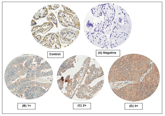
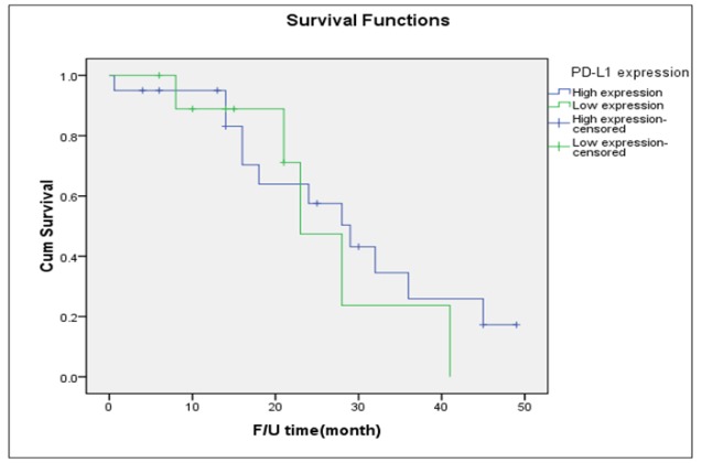
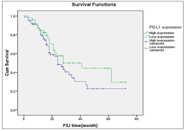
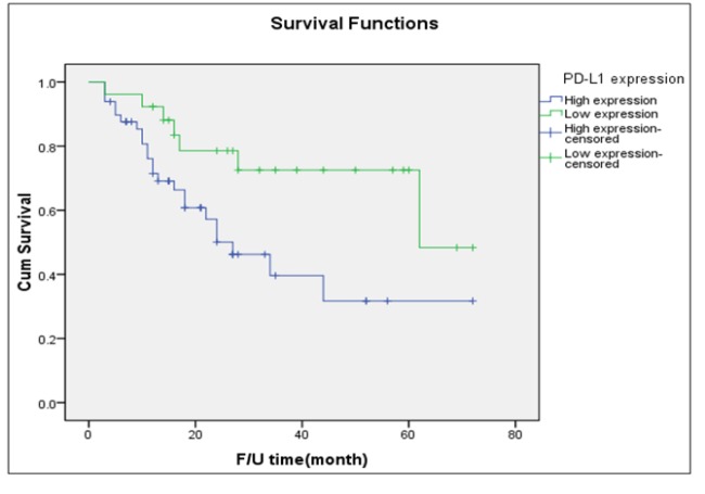
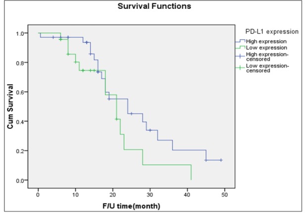
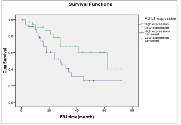
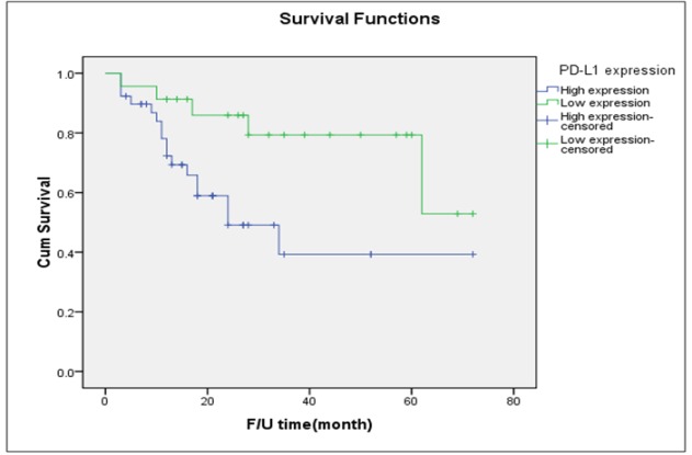
Objective: To examine the expression of programmed death ligand 1 (PD-L1) in type I and type II epithelial ovarian cancers (EOC) and its associations with outcomes. Methods: Records of 132 women with EOC were reviewed. Immunostaining of PD-L1 was performed with formalin-fixed, paraffin-embedded specimens. Expression of PD-L1 was classified into four categories (0; 1+; 2+; 3+) according to intensity of expression. Expression of PD-L1 ≥2+ was deemed to be high. Results: Of the 132 women, 75 (56.8%) and 57 (43.2%) women had type I and type II tumors, respectively. Approximately 70% of cases exhibited high PD-L1 expression. There was no significant difference in the rate of high PD-L1 expression between the two EOC types (65.3% versus 59.6%). In type I tumors, high PD-L1 expression was associated with more advanced stages (51.0% versus 34.6%), greater recurrence (46.9% versus 26.9%), and shorter median progression-free survival (27 months versus 62 months) than low expression. In type II tumors, there were no apparent differences between high and low expression of PD-L1 in terms of the percentage of advanced-stage tumors (82.6% versus 79.4%), recurrence (56.5% versus 58.8%), and median progression-free survival (21 months versus 24 months). Conclusion: high PD-L1 expression is associated with worse oncological outcomes in type I EOC. This finding emphasizes the merit of further studies to confirm this promising result and to determine the potential role of PD-L1 blockade therapy in type I EOC.

摘要

目的

检测程序性死亡配体1(PD-L1)在I型和II型上皮性卵巢癌(EOC)中的表达及其与预后的关系。方法:回顾132例EOC患者的病历。采用福尔马林固定、石蜡包埋标本进行PD-L1免疫染色。根据表达强度将PD-L1表达分为四类(0;1+;2+;3+)。PD-L1表达≥2+被视为高表达。结果:132例患者中,分别有75例(56.8%)和57例(43.2%)患有I型和II型肿瘤。约70%的病例表现为PD-L1高表达。两种EOC类型之间的PD-L1高表达率无显著差异(65.3%对59.6%)。在I型肿瘤中,与低表达相比,PD-L1高表达与更晚期别(51.0%对34.6%)、更高的复发率(46.9%对26.9%)以及更短的无进展生存期中位数(27个月对62个月)相关。在II型肿瘤中,PD-L1高表达和低表达在晚期肿瘤百分比(82.6%对79.4%)、复发率(56.5%对58.8%)以及无进展生存期中位数(21个月对24个月)方面无明显差异。结论:PD-L1高表达与I型EOC更差的肿瘤学预后相关。这一发现强调了进一步研究以证实这一有前景的结果并确定PD-L1阻断治疗在I型EOC中的潜在作用的价值。

https://cdn.ncbi.nlm.nih.gov/pmc/blobs/caef/6948887/60972e2bdc23/APJCP-20-1161-g007.jpg
https://cdn.ncbi.nlm.nih.gov/pmc/blobs/caef/6948887/accb1406b16c/APJCP-20-1161-g001.jpg
https://cdn.ncbi.nlm.nih.gov/pmc/blobs/caef/6948887/331ab07f7607/APJCP-20-1161-g002.jpg
https://cdn.ncbi.nlm.nih.gov/pmc/blobs/caef/6948887/91d90429a7c3/APJCP-20-1161-g003.jpg
https://cdn.ncbi.nlm.nih.gov/pmc/blobs/caef/6948887/3e9fa15b317c/APJCP-20-1161-g004.jpg
https://cdn.ncbi.nlm.nih.gov/pmc/blobs/caef/6948887/8014ca711778/APJCP-20-1161-g005.jpg
https://cdn.ncbi.nlm.nih.gov/pmc/blobs/caef/6948887/978c57521530/APJCP-20-1161-g006.jpg
https://cdn.ncbi.nlm.nih.gov/pmc/blobs/caef/6948887/60972e2bdc23/APJCP-20-1161-g007.jpg

相似文献

1
Programmed Death Ligand 1 (PD-L1) Expression in Epithelial Ovarian Cancer: A Comparison of Type I and Type II Tumors.程序性死亡配体1(PD-L1)在上皮性卵巢癌中的表达:I型和II型肿瘤的比较
Asian Pac J Cancer Prev. 2019 Apr 29;20(4):1161-1169. doi: 10.31557/APJCP.2019.20.4.1161.
2
[Expression and significance of PD-1 and PD-L1 in the specimens of epithelial ovarian cancer].[PD-1和PD-L1在上皮性卵巢癌标本中的表达及意义]
Zhonghua Fu Chan Ke Za Zhi. 2020 Aug 25;55(8):529-534. doi: 10.3760/cma.j.cn112141-20200301-00155.
3
CD105/Ki67 coexpression correlates with tumor progression and poor prognosis in epithelial ovarian cancer.CD105/Ki67 共表达与上皮性卵巢癌的肿瘤进展和不良预后相关。
Int J Gynecol Cancer. 2012 May;22(4):586-92. doi: 10.1097/IGC.0b013e31823c36b8.
4
CA-125 level as a prognostic indicator in type I and type II epithelial ovarian cancer.CA-125 水平作为 I 型和 II 型上皮性卵巢癌的预后指标。
Int J Gynecol Cancer. 2013 Jun;23(5):815-22. doi: 10.1097/IGC.0b013e31828f7a24.
5
Lack of MRE11-RAD50-NBS1 (MRN) complex detection occurs frequently in low-grade epithelial ovarian cancer.在低级别上皮性卵巢癌中,MRE11-RAD50-NBS1(MRN)复合物检测缺失频繁出现。
BMC Cancer. 2017 Jan 10;17(1):44. doi: 10.1186/s12885-016-3026-2.
6
Elevated growth differentiation factor 15 expression predicts poor prognosis in epithelial ovarian cancer patients.生长分化因子15表达升高预示上皮性卵巢癌患者预后不良。
Tumour Biol. 2016 Jul;37(7):9423-31. doi: 10.1007/s13277-015-4699-x. Epub 2016 Jan 18.
7
EphA8 is a prognostic marker for epithelial ovarian cancer.EphA8是上皮性卵巢癌的一个预后标志物。
Oncotarget. 2016 Apr 12;7(15):20801-9. doi: 10.18632/oncotarget.8018.
8
Outcome in serous ovarian cancer is not associated with LATS expression.浆液性卵巢癌的预后与 LATS 表达无关。
J Cancer Res Clin Oncol. 2019 Nov;145(11):2737-2749. doi: 10.1007/s00432-019-03037-4. Epub 2019 Oct 4.
9
Prognostic impact of Dynamin related protein 1 (Drp1) in epithelial ovarian cancer.DRP1 在卵巢上皮性癌中的预后价值。
BMC Cancer. 2020 May 24;20(1):467. doi: 10.1186/s12885-020-06965-4.
10
Periostin in tumor microenvironment is associated with poor prognosis and platinum resistance in epithelial ovarian carcinoma.肿瘤微环境中的骨膜蛋白与上皮性卵巢癌的不良预后和铂耐药相关。
Oncotarget. 2016 Jan 26;7(4):4036-47. doi: 10.18632/oncotarget.6700.

引用本文的文献

1
Comparison of PD-L1, CTR-1, VEGF, and p53 expression in sensitive and resistant epithelial ovarian cancer (EOC) patients to platinum-based chemotherapy.铂类化疗敏感和耐药的上皮性卵巢癌(EOC)患者中PD-L1、CTR-1、VEGF和p53表达的比较。
Narra J. 2025 Apr;5(1):e1419. doi: 10.52225/narra.v5i1.1419. Epub 2025 Jan 6.
2
Correlation of PD-L1 expression with different clinico-pathological and immunohistochemical features of ovarian surface epithelial tumors.PD-L1表达与卵巢表面上皮性肿瘤不同临床病理及免疫组化特征的相关性
Clin Transl Oncol. 2025 Feb;27(2):699-705. doi: 10.1007/s12094-024-03613-2. Epub 2024 Aug 1.
3

本文引用的文献

1
The prognostic effect of neoadjuvant chemoradiotherapy on the change of PD-L1 expression in patients with locally advanced rectal adenocarcinoma.新辅助放化疗对局部晚期直肠腺癌患者PD-L1表达变化的预后影响
J BUON. 2017 Jul-Aug;22(4):875-881.
2
Prognostic value of programmed death-ligand 1 (PD-L1) expression in ovarian clear cell carcinoma.程序性死亡配体1(PD-L1)表达在卵巢透明细胞癌中的预后价值
J Gynecol Oncol. 2017 Nov;28(6):e77. doi: 10.3802/jgo.2017.28.e77.
3
PD-1 and PD-L1 Checkpoint Signaling Inhibition for Cancer Immunotherapy: Mechanism, Combinations, and Clinical Outcome.
Mesenchymal ovarian cancer cells promote CD8 T cell exhaustion through the LGALS3-LAG3 axis.
间充质卵巢癌细胞通过 LGALS3-LAG3 轴促进 CD8 T 细胞衰竭。
NPJ Syst Biol Appl. 2023 Dec 12;9(1):61. doi: 10.1038/s41540-023-00322-4.
4
Low plasma PD-L1 levels, early tumor onset and absence of peritoneal carcinomatosis improve prognosis of women with advanced high-grade serous ovarian cancer.低血浆 PD-L1 水平、早期肿瘤发生和无腹膜癌转移可改善晚期高级别浆液性卵巢癌女性的预后。
BMC Cancer. 2023 May 13;23(1):437. doi: 10.1186/s12885-023-10911-5.
5
Focus on PD-1/PD-L1 as a Therapeutic Target in Ovarian Cancer.关注 PD-1/PD-L1 作为卵巢癌的治疗靶点。
Int J Mol Sci. 2022 Oct 11;23(20):12067. doi: 10.3390/ijms232012067.
6
PD-L1 Expression in Different Segments and Histological Types of Ovarian Cancer According to Lymphocytic Infiltrate.根据淋巴细胞浸润情况分析卵巢癌不同节段和组织学类型的 PD-L1 表达
Medicina (Kaunas). 2021 Nov 29;57(12):1309. doi: 10.3390/medicina57121309.
7
Clinical and Prognostic Value of Antigen-Presenting Cells with PD-L1/PD-L2 Expression in Ovarian Cancer Patients.卵巢癌患者 PD-L1/PD-L2 表达的抗原呈递细胞的临床和预后价值。
Int J Mol Sci. 2021 Oct 26;22(21):11563. doi: 10.3390/ijms222111563.
8
Predictive Values of Programmed Cell Death-Ligand 1 Expression for Prognosis, Clinicopathological Factors, and Response to Programmed Cell Death-1/Programmed Cell Death-Ligand 1 Inhibitors in Patients With Gynecological Cancers: A Meta-Analysis.程序性细胞死亡配体1表达对妇科癌症患者预后、临床病理因素及程序性细胞死亡蛋白1/程序性细胞死亡配体1抑制剂反应的预测价值:一项荟萃分析
Front Oncol. 2021 Feb 1;10:572203. doi: 10.3389/fonc.2020.572203. eCollection 2020.
9
Hypoxia-Mediated Decrease of Ovarian Cancer Cells Reaction to Treatment: Significance for Chemo- and Immunotherapies.缺氧介导的卵巢癌细胞对治疗反应的降低:对化疗和免疫治疗的意义。
Int J Mol Sci. 2020 Dec 14;21(24):9492. doi: 10.3390/ijms21249492.
10
Endometriosis-Associated Massive Ascites in an Asian Woman: A Case Report of a Rare Clinical Entity.一名亚洲女性的子宫内膜异位症相关性大量腹水:一种罕见临床病症的病例报告
Case Rep Obstet Gynecol. 2020 Aug 4;2020:8879643. doi: 10.1155/2020/8879643. eCollection 2020.
用于癌症免疫治疗的PD-1和PD-L1检查点信号抑制:机制、联合应用及临床结果
Front Pharmacol. 2017 Aug 23;8:561. doi: 10.3389/fphar.2017.00561. eCollection 2017.
4
Prognostic value of tumor PD-L1 expression combined with CD8 tumor infiltrating lymphocytes in high grade serous ovarian cancer.肿瘤 PD-L1 表达联合 CD8 肿瘤浸润淋巴细胞对高级别浆液性卵巢癌的预后价值。
Int Immunopharmacol. 2017 Nov;52:7-14. doi: 10.1016/j.intimp.2017.08.017. Epub 2017 Aug 31.
5
Effect of neoadjuvant chemotherapy on tumor-infiltrating lymphocytes and PD-L1 expression in breast cancer and its clinical significance.新辅助化疗对乳腺癌肿瘤浸润淋巴细胞及PD-L1表达的影响及其临床意义
Breast Cancer Res. 2017 Aug 7;19(1):91. doi: 10.1186/s13058-017-0884-8.
6
Immune contexture and histological response after neoadjuvant chemotherapy predict clinical outcome of lung cancer patients.新辅助化疗后的免疫微环境和组织学反应可预测肺癌患者的临床结局。
Oncoimmunology. 2016 Dec 8;5(12):e1255394. doi: 10.1080/2162402X.2016.1255394. eCollection 2016.
7
Neoadjuvant chemotherapy (NACT) increases immune infiltration and programmed death-ligand 1 (PD-L1) expression in epithelial ovarian cancer (EOC).新辅助化疗(NACT)增加上皮性卵巢癌(EOC)中的免疫浸润和程序性死亡配体 1(PD-L1)表达。
Ann Oncol. 2017 Mar 1;28(3):651-657. doi: 10.1093/annonc/mdw625.
8
Altered expression of programmed death-ligand 1 after neo-adjuvant chemotherapy in patients with lung squamous cell carcinoma.肺鳞状细胞癌患者新辅助化疗后程序性死亡配体1表达的改变
Lung Cancer. 2016 Sep;99:166-71. doi: 10.1016/j.lungcan.2016.07.013. Epub 2016 Jul 15.
9
Neoadjuvant Chemotherapy Modulates the Immune Microenvironment in Metastases of Tubo-Ovarian High-Grade Serous Carcinoma.新辅助化疗调节卵巢高级别浆液性癌转移灶的免疫微环境。
Clin Cancer Res. 2016 Jun 15;22(12):3025-36. doi: 10.1158/1078-0432.CCR-15-2657.
10
The significance and therapeutic potential of PD-1 and its ligands in ovarian cancer: A systematic review.PD-1及其配体在卵巢癌中的意义及治疗潜力:一项系统综述。
Gynecol Oncol. 2016 Jul;142(1):184-189. doi: 10.1016/j.ygyno.2016.04.002. Epub 2016 Apr 15.