Weld Alistair, Dixon Luke, Anichini Giulio, Patel Neekhil, Nimer Amr, Dyck Michael, O'Neill Kevin, Lim Adrian, Giannarou Stamatia, Camp Sophie
Hamlyn Centre, Imperial College London, Exhibition Rd, London, SW7 2AZ, UK.
Department of Imaging, Charing Cross Hospital, Fulham Palace Rd, London, W6 8RF, UK.
Acta Neurochir (Wien). 2024 Aug 1;166(1):317. doi: 10.1007/s00701-024-06179-8.
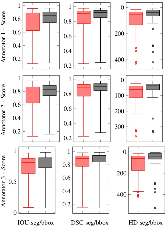
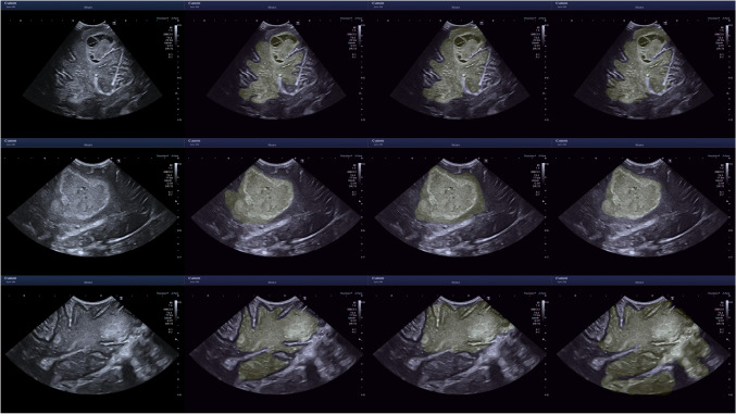
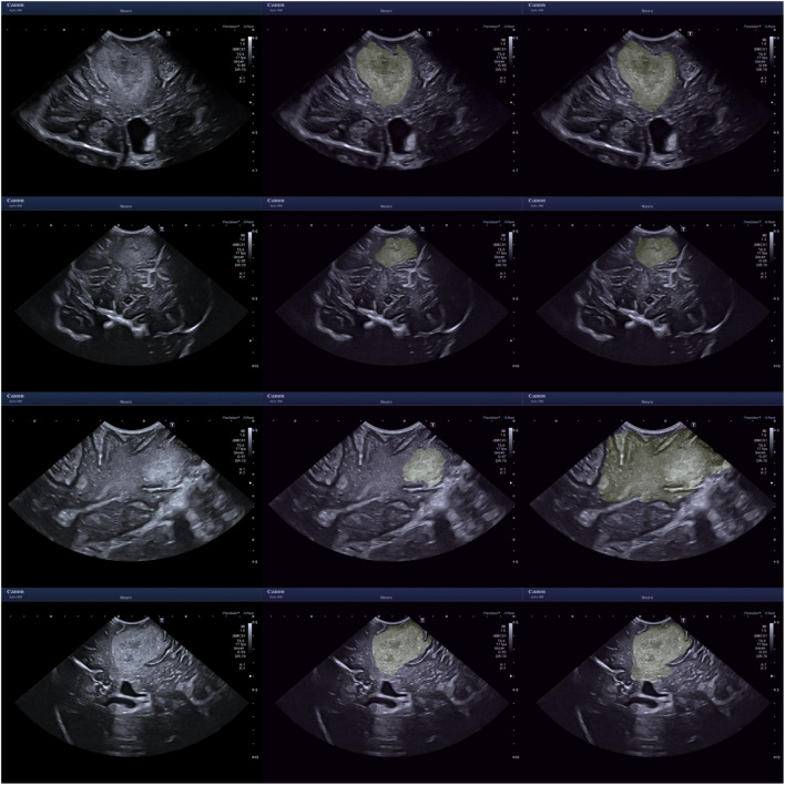
Objective - Addressing the challenges that come with identifying and delineating brain tumours in intraoperative ultrasound. Our goal is to both qualitatively and quantitatively assess the interobserver variation, amongst experienced neuro-oncological intraoperative ultrasound users (neurosurgeons and neuroradiologists), in detecting and segmenting brain tumours on ultrasound. We then propose that, due to the inherent challenges of this task, annotation by localisation of the entire tumour mass with a bounding box could serve as an ancillary solution to segmentation for clinical training, encompassing margin uncertainty and the curation of large datasets. Methods - 30 ultrasound images of brain lesions in 30 patients were annotated by 4 annotators - 1 neuroradiologist and 3 neurosurgeons. The annotation variation of the 3 neurosurgeons was first measured, and then the annotations of each neurosurgeon were individually compared to the neuroradiologist's, which served as a reference standard as their segmentations were further refined by cross-reference to the preoperative magnetic resonance imaging (MRI). The following statistical metrics were used: Intersection Over Union (IoU), Sørensen-Dice Similarity Coefficient (DSC) and Hausdorff Distance (HD). These annotations were then converted into bounding boxes for the same evaluation. Results - There was a moderate level of interobserver variance between the neurosurgeons and a larger level of variance when compared against the MRI-informed reference standard annotations by the neuroradiologist, mean across annotators . After converting the segments to bounding boxes, all metrics improve, most significantly, the interquartile range drops by . Conclusion - This study highlights the current challenges with detecting and defining tumour boundaries in neuro-oncological intraoperative brain ultrasound. We then show that bounding box annotation could serve as a useful complementary approach for both clinical and technical reasons.
目的——应对术中超声识别和勾勒脑肿瘤所带来的挑战。我们的目标是定性和定量评估经验丰富的神经肿瘤术中超声使用者(神经外科医生和神经放射科医生)在超声检测和分割脑肿瘤时的观察者间差异。然后我们提出,由于这项任务存在固有挑战,用边界框定位整个肿瘤块进行标注可作为临床训练分割的辅助解决方案,涵盖边界不确定性和大型数据集的整理。方法——4名标注人员(1名神经放射科医生和3名神经外科医生)对30例患者的30张脑病变超声图像进行标注。首先测量3名神经外科医生的标注差异,然后将每名神经外科医生的标注分别与神经放射科医生的标注进行比较,神经放射科医生的标注作为参考标准,因为他们的分割通过与术前磁共振成像(MRI)交叉参考得到了进一步完善。使用了以下统计指标:交并比(IoU)、索伦森-戴斯相似系数(DSC)和豪斯多夫距离(HD)。然后将这些标注转换为边界框进行相同评估。结果——神经外科医生之间存在中等程度的观察者间差异,与神经放射科医生基于MRI的参考标准标注相比差异更大,所有标注人员的平均值 。将分割结果转换为边界框后,所有指标均有所改善,最显著的是四分位间距下降了 。结论——本研究突出了神经肿瘤术中脑超声检测和定义肿瘤边界目前面临的挑战。然后我们表明,出于临床和技术原因,边界框标注可作为一种有用的补充方法。