Universidade Federal de Pelotas - UFPel, Dental School, Diagnostic Center for Oral Diseases, Pelotas, RS, Brazil.
Universidade Estadual de Campinas - Unicamp, Piracicaba Dental School, Department of Oral Diagnosis, Piracicaba, SP, Brazil.
Braz Oral Res. 2024 Aug 5;38:e069. doi: 10.1590/1807-3107bor-2024.vol38.0069. eCollection 2024.
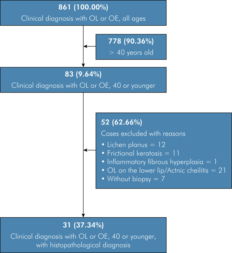
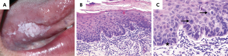
The objective of the present study was to investigate the frequency of oral leukoplakia and oral erythroplakia among young patients from three Brazilian reference centers in Oral and Maxillofacial Pathology. A retrospective study was carried out from 2011 to 2021 on 861 patients diagnosed with oral leukoplakia and oral erythroplakia. Demographic and clinicopathological data were evaluated. Fisher's exact test was used to evaluate the association among sex, age, anatomical location, and histopathological diagnosis. A total of 83 (9.64%) cases involved young patients (aged <40 years). Among these, biopsy records were included in 31 (37.34%) cases, all of which received a clinical diagnosis of oral leukoplakia. Seventeen (54.84%) patients were female, mostly in their fourth decade of life (n = 22/70.97%), and their mean age at diagnosis was 32.61(± 5.21) years. Among informed cases, seven (22.58%) patients were smokers. The lateral border of the tongue (n = 9/29.03%) was the most affected site. In 13 (41.94%) cases, oral leukoplakias showed a homogeneous appearance. The mean size of the lesions was 1.47 cm (0.2-3.0 cm) and the mean time of disease progression was 64.37 (± 65.90) months. The histopathological analysis showed that 11 cases (35.48%) exhibited some degree of epithelial dysplasia. Acanthosis and/or hyperkeratosis were observed in 20 cases (64.52%). No significant associations were observed between sex and anatomical location, age and anatomical location, nor between sex and histological diagnosis (p > 0.05). Oral leukoplakia and oral erythroplakia are uncommon diseases in young patients. In this population, oral leukoplakia shows a slight predilection for women aged between 30 and 39 years.
本研究的目的是调查三家巴西口腔颌面病理学参考中心的年轻患者中口腔白斑病和口腔红斑病的频率。对 2011 年至 2021 年期间诊断为口腔白斑病和口腔红斑病的 861 例患者进行了回顾性研究。评估了人口统计学和临床病理学数据。Fisher 确切检验用于评估性别、年龄、解剖部位和组织病理学诊断之间的关联。共有 83 例(9.64%)患者为年轻患者(<40 岁)。其中,31 例(37.34%)患者有活检记录,均被临床诊断为口腔白斑病。17 例(54.84%)患者为女性,年龄主要集中在第四个十年(n = 22/70.97%),诊断时的平均年龄为 32.61(±5.21)岁。在知情患者中,有 7 例(22.58%)患者为吸烟者。受影响最严重的部位是舌侧缘(n = 9/29.03%)。在 13 例(41.94%)口腔白斑病患者中,病变表现为均质外观。病变的平均大小为 1.47cm(0.2-3.0cm),疾病进展的平均时间为 64.37(±65.90)个月。组织病理学分析显示 11 例(35.48%)存在一定程度的上皮异型增生。20 例(64.52%)可见棘层肥厚和/或过度角化。性别和解剖部位、年龄和解剖部位以及性别和组织学诊断之间无显著相关性(p > 0.05)。口腔白斑病和口腔红斑病在年轻患者中并不常见。在该人群中,口腔白斑病女性患者略多于男性,年龄在 30-39 岁之间。