Fukuda Yosuke, Ishikawa Keijiro, Kiyohara Kohei, Maehara Yusuke, Ji Rui, Mori Kenichiro, Kobayashi Yoshiyuki, Akiyama Masato, Nakama Takahito, Notomi Shoji, Shiose Satomi, Takeda Atsunobu, Sonoda Koh-Hei
Department of Ophthalmology, Graduate School of Medical Sciences, Kyushu University, Fukuoka, Japan.
Department of Ophthalmology, Aso Iizuka Hospital, Fukuoka, Japan.
Transl Vis Sci Technol. 2024 Aug 1;13(8):13. doi: 10.1167/tvst.13.8.13.
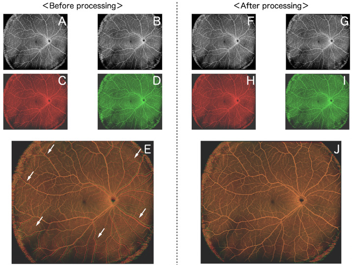
Proliferative retinal changes may occur postsurgery for rhegmatogenous retinal detachment (RRD), possibly preceding recurrent detachment. This study aims to establish the groundwork for an imaging system capable of discerning changes in retinal vessel tortuosity after RRD repair, analyzing widefield optical coherence tomography angiography (WF-OCTA) images.
Eighty-eight eyes of 86 patients with RRD who underwent surgical procedures and had repeated imaging with clear widefield optical coherence tomography (WF-OCT) and WF-OCTA on different postoperative days were enrolled in this retrospective study. We compared WF-OCTA images over time to identify alterations in retinal vessel tortuosity and observed regional changes in retinal morphology.
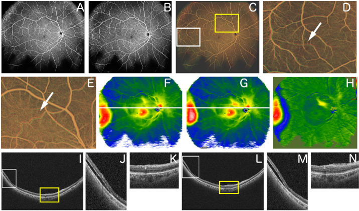
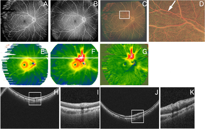
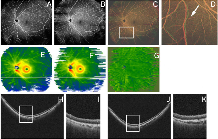
After image processing, changes in retinal vessel tortuosity were detected in 66 quadrants. These changes, attributed to retinal traction from proliferative membranes, were observed in 56 quadrants, among which retinal thickness remained unchanged in seven sectors (12.5%) according to the WF-OCT map. In nine quadrants, changes in retinal vessel tortuosity were attributed to changes in subretinal fluid, aligning with observable variations in retinal thickness.
Observation of vessel tortuosity changes using WF-OCTA can help detect early postoperative proliferative changes in eyes with RRD.
Because WF-OCTA can detect minute vessel tortuosity changes, it can offer a noninvasive alternative for the detection of early postoperative proliferative changes.
孔源性视网膜脱离(RRD)手术后可能会出现视网膜增殖性改变,这可能先于复发性视网膜脱离。本研究旨在为一种能够识别RRD修复后视网膜血管迂曲变化的成像系统奠定基础,对超广角光学相干断层扫描血管造影(WF-OCTA)图像进行分析。
本回顾性研究纳入了86例接受手术治疗的RRD患者的88只眼,这些患者在术后不同时间进行了清晰的超广角光学相干断层扫描(WF-OCT)和WF-OCTA重复成像。我们比较不同时间的WF-OCTA图像,以识别视网膜血管迂曲的变化,并观察视网膜形态的区域变化。
经过图像处理,在66个象限中检测到视网膜血管迂曲的变化。这些变化归因于增殖膜引起的视网膜牵拉,在56个象限中观察到,根据WF-OCT图,其中7个扇区(12.5%)的视网膜厚度保持不变。在9个象限中,视网膜血管迂曲的变化归因于视网膜下液的变化,与视网膜厚度的可观察到的变化一致。
使用WF-OCTA观察血管迂曲变化有助于检测RRD患者术后早期的增殖性变化。
由于WF-OCTA可以检测到微小的血管迂曲变化,它可以为检测术后早期增殖性变化提供一种非侵入性的替代方法。