• 文献检索
  • 文档翻译
  • 深度研究
  • 学术资讯
  • Suppr Zotero 插件Zotero 插件
  • 邀请有礼
  • 套餐&价格
  • 历史记录
应用&插件
Suppr Zotero 插件Zotero 插件浏览器插件Mac 客户端Windows 客户端微信小程序
定价
高级版会员购买积分包购买API积分包
服务
文献检索文档翻译深度研究API 文档MCP 服务
关于我们
关于 Suppr公司介绍联系我们用户协议隐私条款
关注我们

Suppr 超能文献

核心技术专利:CN118964589B侵权必究
粤ICP备2023148730 号-1Suppr @ 2026

文献检索

告别复杂PubMed语法,用中文像聊天一样搜索,搜遍4000万医学文献。AI智能推荐,让科研检索更轻松。

立即免费搜索

文件翻译

保留排版,准确专业,支持PDF/Word/PPT等文件格式,支持 12+语言互译。

免费翻译文档

深度研究

AI帮你快速写综述,25分钟生成高质量综述,智能提取关键信息,辅助科研写作。

立即免费体验

用于研究核结构和基因表达的高通量图像处理软件。

High-throughput image processing software for the study of nuclear architecture and gene expression.

作者信息

Keikhosravi Adib, Almansour Faisal, Bohrer Christopher H, Fursova Nadezda A, Guin Krishnendu, Sood Varun, Misteli Tom, Larson Daniel R, Pegoraro Gianluca

机构信息

High-Throughput Imaging Facility, National Cancer Institute, National Institute of Health, Bethesda, MD, 20892, USA.

Cell Biology of Genomes, National Cancer Institute, National Institute of Health, Bethesda, MD, 20892, USA.

出版信息

Sci Rep. 2024 Aug 8;14(1):18426. doi: 10.1038/s41598-024-66600-1.

DOI:10.1038/s41598-024-66600-1
PMID:39117696
原文链接:https://pmc.ncbi.nlm.nih.gov/articles/PMC11310328/
Abstract

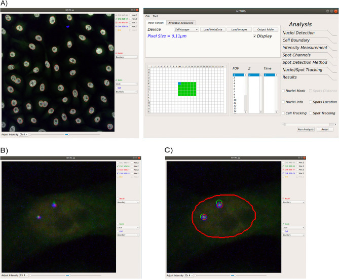
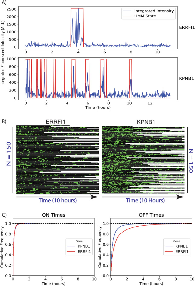
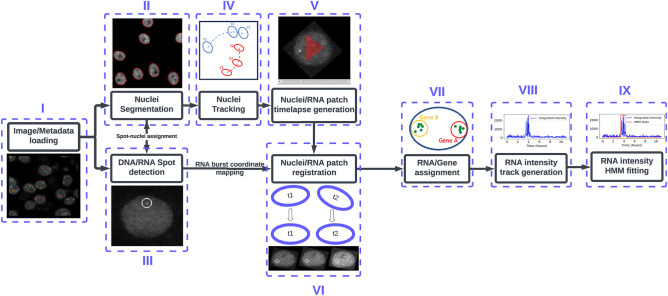
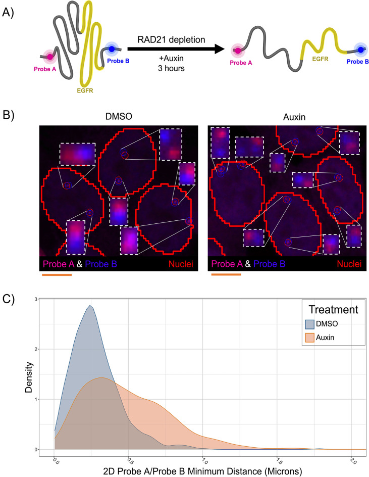
High-throughput imaging (HTI) generates complex imaging datasets from a large number of experimental perturbations. Commercial HTI software programs for image analysis workflows typically do not allow full customization and adoption of new image processing algorithms in the analysis modules. While open-source HTI analysis platforms provide individual modules in the workflow, like nuclei segmentation, spot detection, or cell tracking, they are often limited in integrating novel analysis modules or algorithms. Here, we introduce the High-Throughput Image Processing Software (HiTIPS) to expand the range and customization of existing HTI analysis capabilities. HiTIPS incorporates advanced image processing and machine learning algorithms for automated cell and nuclei segmentation, spot signal detection, nucleus tracking, nucleus registration, spot tracking, and quantification of spot signal intensity. Furthermore, HiTIPS features a graphical user interface that is open to integration of new analysis modules for existing analysis pipelines and to adding new analysis modules. To demonstrate the utility of HiTIPS, we present three examples of image analysis workflows for high-throughput DNA FISH, immunofluorescence (IF), and live-cell imaging of transcription in single cells. Altogether, we demonstrate that HiTIPS is a user-friendly, flexible, and open-source HTI software platform for a variety of cell biology applications.

摘要

高通量成像(HTI)通过大量实验扰动生成复杂的成像数据集。用于图像分析工作流程的商业HTI软件程序通常不允许在分析模块中进行完全定制和采用新的图像处理算法。虽然开源HTI分析平台在工作流程中提供了诸如细胞核分割、斑点检测或细胞追踪等单个模块,但它们在集成新的分析模块或算法方面往往受到限制。在此,我们引入高通量图像处理软件(HiTIPS)以扩展现有HTI分析能力的范围和定制性。HiTIPS整合了先进的图像处理和机器学习算法,用于自动细胞和细胞核分割、斑点信号检测、细胞核追踪、细胞核配准、斑点追踪以及斑点信号强度定量。此外,HiTIPS具有图形用户界面,可用于为现有分析流程集成新的分析模块以及添加新的分析模块。为证明HiTIPS的实用性,我们展示了高通量DNA荧光原位杂交(FISH)、免疫荧光(IF)以及单细胞转录活细胞成像的三个图像分析工作流程示例。总之,我们证明HiTIPS是一个适用于多种细胞生物学应用的用户友好、灵活且开源的HTI软件平台。

https://cdn.ncbi.nlm.nih.gov/pmc/blobs/1158/11310328/5b0364d31ac5/41598_2024_66600_Fig5_HTML.jpg
https://cdn.ncbi.nlm.nih.gov/pmc/blobs/1158/11310328/89792dba5a8e/41598_2024_66600_Fig1_HTML.jpg
https://cdn.ncbi.nlm.nih.gov/pmc/blobs/1158/11310328/d26d7aa14252/41598_2024_66600_Fig2_HTML.jpg
https://cdn.ncbi.nlm.nih.gov/pmc/blobs/1158/11310328/fe86eaf004a6/41598_2024_66600_Fig3_HTML.jpg
https://cdn.ncbi.nlm.nih.gov/pmc/blobs/1158/11310328/194ea123c91e/41598_2024_66600_Fig4_HTML.jpg
https://cdn.ncbi.nlm.nih.gov/pmc/blobs/1158/11310328/5b0364d31ac5/41598_2024_66600_Fig5_HTML.jpg

相似文献

1
High-throughput image processing software for the study of nuclear architecture and gene expression.用于研究核结构和基因表达的高通量图像处理软件。
Sci Rep. 2024 Aug 8;14(1):18426. doi: 10.1038/s41598-024-66600-1.
2
HiTIPS: High-Throughput Image Processing Software for the Study of Nuclear Architecture and Gene Expression.HiTIPS:用于细胞核结构与基因表达研究的高通量图像处理软件。
bioRxiv. 2023 Nov 5:2023.11.02.565366. doi: 10.1101/2023.11.02.565366.
3
Multi-objective Parameter Auto-tuning for Tissue Image Segmentation Workflows.用于组织图像分割工作流程的多目标参数自动调整
J Digit Imaging. 2019 Jun;32(3):521-533. doi: 10.1007/s10278-018-0138-z.
4
Image analysis workflows to reveal the spatial organization of cell nuclei and chromosomes.图像分析工作流程揭示细胞核和染色体的空间组织。
Nucleus. 2022 Dec;13(1):277-299. doi: 10.1080/19491034.2022.2144013.
5
DolphinNext: a distributed data processing platform for high throughput genomics.海豚下一代:一个用于高通量基因组学的分布式数据处理平台。
BMC Genomics. 2020 Apr 19;21(1):310. doi: 10.1186/s12864-020-6714-x.
6
FocAn: automated 3D analysis of DNA repair foci in image stacks acquired by confocal fluorescence microscopy.FocAn:通过共聚焦荧光显微镜获取的图像堆栈中 DNA 修复焦点的自动 3D 分析。
BMC Bioinformatics. 2020 Jan 28;21(1):27. doi: 10.1186/s12859-020-3370-8.
7
CellAnimation: an open source MATLAB framework for microscopy assays.细胞动画:一个用于显微镜检测的开源 MATLAB 框架。
Bioinformatics. 2012 Jan 1;28(1):138-9. doi: 10.1093/bioinformatics/btr633. Epub 2011 Nov 24.
8
Smart 3D-FISH: automation of distance analysis in nuclei of interphase cells by image processing.智能三维荧光原位杂交技术:通过图像处理实现间期细胞核距离分析的自动化
Cytometry A. 2005 Sep;67(1):18-26. doi: 10.1002/cyto.a.20170.
9
An open-source solution for advanced imaging flow cytometry data analysis using machine learning.一种使用机器学习进行高级成像流式细胞术数据分析的开源解决方案。
Methods. 2017 Jan 1;112:201-210. doi: 10.1016/j.ymeth.2016.08.018. Epub 2016 Sep 2.
10
Oufti: an integrated software package for high-accuracy, high-throughput quantitative microscopy analysis.Oufti:一款用于高精度、高通量定量显微镜分析的集成软件包。
Mol Microbiol. 2016 Feb;99(4):767-77. doi: 10.1111/mmi.13264. Epub 2015 Dec 18.

引用本文的文献

1
Orderly mitosis shapes interphase genome architecture.有序的有丝分裂塑造了间期基因组结构。
bioRxiv. 2025 Jun 3:2025.06.03.657645. doi: 10.1101/2025.06.03.657645.
2
Simulation and Quantitative Analysis of Spatial Centromere Distribution Patterns.着丝粒空间分布模式的模拟与定量分析
Cells. 2025 Mar 25;14(7):491. doi: 10.3390/cells14070491.
3
Simulation and quantitative analysis of spatial centromere distribution patterns.着丝粒空间分布模式的模拟与定量分析

本文引用的文献

1
High-throughput Oligopaint screen identifies druggable 3D genome regulators.高通量寡探针筛选鉴定可成药的 3D 基因组调控因子
Nature. 2023 Aug;620(7972):209-217. doi: 10.1038/s41586-023-06340-w. Epub 2023 Jul 12.
2
OME-Zarr: a cloud-optimized bioimaging file format with international community support.OME-Zarr:具有国际社区支持的云优化生物成像文件格式。
Histochem Cell Biol. 2023 Sep;160(3):223-251. doi: 10.1007/s00418-023-02209-1. Epub 2023 Jul 10.
3
Identification of epigenetic modulators as determinants of nuclear size and shape.
bioRxiv. 2025 Jan 24:2025.01.22.634320. doi: 10.1101/2025.01.22.634320.
4
Mechano-osmotic signals control chromatin state and fate transitions in pluripotent stem cells.机械渗透信号控制多能干细胞中的染色质状态和命运转变。
bioRxiv. 2024 Sep 7:2024.09.07.611779. doi: 10.1101/2024.09.07.611779.
5
Allele-level visualization of transcription and chromatin by high-throughput imaging.通过高通量成像对转录和染色质进行等位基因水平的可视化。
bioRxiv. 2024 Feb 19:2024.02.19.580973. doi: 10.1101/2024.02.19.580973.
6
Allele-level visualization of transcription and chromatin by high-throughput imaging.通过高通量成像实现转录和染色质的等位基因水平可视化。
Res Sq. 2024 Feb 22:rs.3.rs-3970096. doi: 10.21203/rs.3.rs-3970096/v1.
鉴定表观遗传调节剂作为核大小和形状的决定因素。
Elife. 2023 May 23;12:e80653. doi: 10.7554/eLife.80653.
4
MoBIE: a Fiji plugin for sharing and exploration of multi-modal cloud-hosted big image data.MoBIE:一款用于共享和探索多模态云托管大图像数据的斐济插件。
Nat Methods. 2023 Apr;20(4):475-476. doi: 10.1038/s41592-023-01776-4.
5
RS-FISH: precise, interactive, fast, and scalable FISH spot detection.RS-FISH:精确、交互、快速且可扩展的 FISH 斑点检测。
Nat Methods. 2022 Dec;19(12):1563-1567. doi: 10.1038/s41592-022-01669-y. Epub 2022 Nov 17.
6
Cellpose 2.0: how to train your own model.Cellpose 2.0:如何训练自己的模型。
Nat Methods. 2022 Dec;19(12):1634-1641. doi: 10.1038/s41592-022-01663-4. Epub 2022 Nov 7.
7
High-Throughput DNA FISH (hiFISH).高通量 DNA 荧光原位杂交(hiFISH)。
Methods Mol Biol. 2022;2532:245-274. doi: 10.1007/978-1-0716-2497-5_12.
8
GIANI - open-source software for automated analysis of 3D microscopy images.GIANI - 用于 3D 显微镜图像自动分析的开源软件。
J Cell Sci. 2022 May 15;135(10). doi: 10.1242/jcs.259511. Epub 2022 May 17.
9
Interrogating RNA and protein spatial subcellular distribution in smFISH data with DypFISH.使用 DypFISH 对 smFISH 数据中的 RNA 和蛋白质的空间亚细胞分布进行检测。
Cell Rep Methods. 2021 Sep 13;1(5):100068. doi: 10.1016/j.crmeth.2021.100068. eCollection 2021 Sep 27.
10
FISH-quant v2: a scalable and modular tool for smFISH image analysis.FISH-quant v2:一种用于 smFISH 图像分析的可扩展和模块化工具。
RNA. 2022 Jun;28(6):786-795. doi: 10.1261/rna.079073.121. Epub 2022 Mar 28.