Faculty of Dentistry, Department of Prosthodontics, Gaziantep University, Gaziantep, Turkey.
Faculty of medicine, Department of medical microbiology, Sanko University, Gaziantep, Turkey.
Lasers Med Sci. 2024 Aug 9;39(1):212. doi: 10.1007/s10103-024-04166-0.
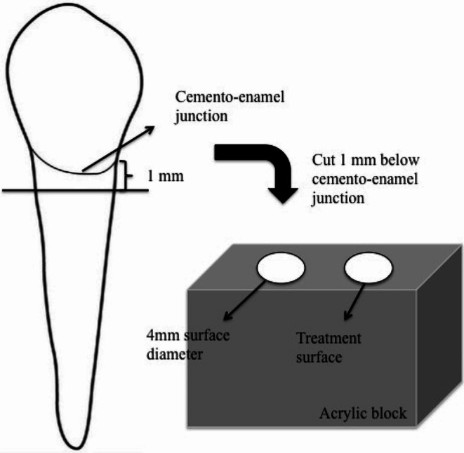
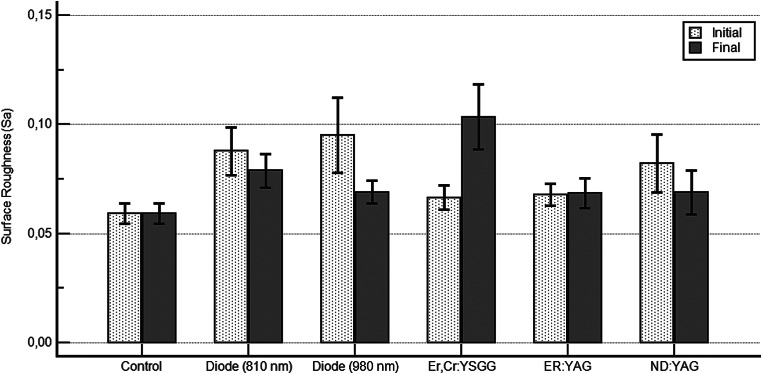
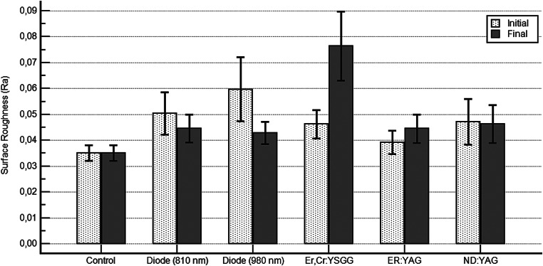
The aim of the study was to measure the degree of dentine surface roughness caused by five distinct lasers used to treat dentine hypersensitivity, as well as to evaluate the subsequent bacterial colonization on these irradiated surfaces. Sixty human maxillary premolar teeth without caries or restoration which were extracted for periodontal reasons were used in this study. Five different types of lasers were applied to the root dentin surface. Tested samples were divided into six groups of 10 samples each; control, diode (810 nm), diode (980 nm), Nd: YAG, Er: YAG, and Er, Cr: YSGG laser groups. The arithmetic mean of the surface roughness values (Ra) and the average roughness over a measurement area (Sa) were measured pre- and post-application using any of the laser types. Swab samples were then collected from the dentin surface. Following a 24-hour incubation period at 37 °C, the colony forming units were counted using a stereoscope. The results demonstrated a statistically significant difference in the surface roughness values pre- and post-application (Ra and Sa, respectively) in the Er, Cr: YSGG laser group (p = 0.037,p = 0.007). No significant difference was observed in the other groups (p > 0.05). There was no statistically significant difference in the number of bacterial colonies observed between the test and control groups. Diode and Nd: YAG lasers showed either a decrease or no change in surface roughness; however, the hard tissue lasers (Er: YAG, Er, Cr: YSGG) showed an increase. The Er: YAG and Nd: YAG laser groups exhibited decreased bacterial adhesion compared to the other groups.
本研究旨在测量五种不同激光治疗牙本质敏感症时引起的牙本质表面粗糙度程度,并评估这些辐照表面随后的细菌定植。本研究使用了 60 颗因牙周原因而拔除的无龋或修复的上颌前磨牙。将五种不同类型的激光应用于牙根牙本质表面。测试样本分为 6 组,每组 10 个样本;对照组、二极管(810nm)、二极管(980nm)、Nd:YAG、Er:YAG 和 Er、Cr:YSGG 激光组。使用任何一种激光类型测量处理前后表面粗糙度值(Ra)和测量区域平均粗糙度(Sa)的算术平均值。然后从牙本质表面采集拭子样本。在 37°C 孵育 24 小时后,使用立体显微镜计数集落形成单位。结果表明,Er、Cr:YSGG 激光组处理前后表面粗糙度值(Ra 和 Sa)存在统计学差异(p=0.037,p=0.007)。其他组无统计学差异(p>0.05)。在测试组和对照组之间,观察到的细菌菌落数量没有统计学差异。二极管和 Nd:YAG 激光显示表面粗糙度降低或不变;然而,硬组织激光(Er:YAG、Er、Cr:YSGG)则显示出增加。与其他组相比,Er:YAG 和 Nd:YAG 激光组的细菌黏附减少。