Ibrahim Ibrahim, Škoch Antonín, Dezortová Monika, Adla Theodor, Flusserová Vlasta, Nagy Markéta, Douchová Irena, Fialová Martina, Filová Vanda, Pajuelo Dita, Ibrahimová Markéta, Tintěra Jaroslav
MR Unit, Department of Diagnostic and Interventional Radiology, Institute for Clinical and Experimental Medicine, Prague, Czech Republic.
Specialised Outpatient Care Division, Department of Neurology, Institute for Clinical and Experimental Medicine, Prague, Czech Republic.
Quant Imaging Med Surg. 2024 Aug 1;14(8):5499-5512. doi: 10.21037/qims-24-162. Epub 2024 Jul 12.
Changes in both the vascular system and brain tissues can occur after a prior episode of coronavirus disease 2019 (COVID-19), detectable through modifications in diffusion parameters using magnetic resonance imaging (MRI) techniques. These changes in diffusion parameters may be particularly prominent in highly organized structures such as the corpus callosum (CC), including its major components, which have not been adequately studied following COVID-19 infection. Therefore, the study aimed to evaluate microstructural changes in whole-brain (WB) diffusion, with a specific focus on the CC.
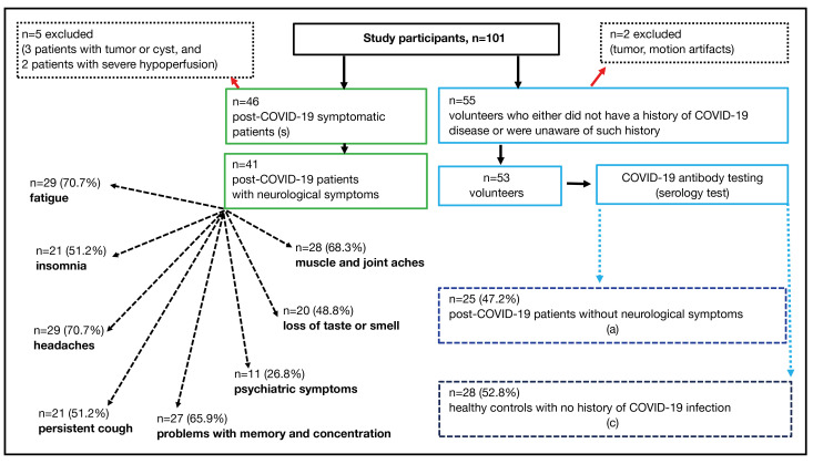
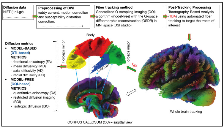
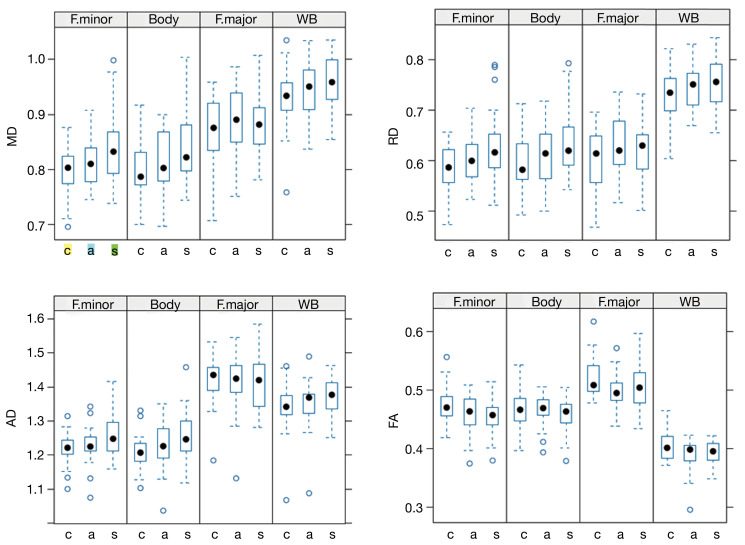
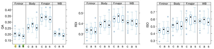
A total of 101 probands (age range from 18 to 69 years) participated in this retrospective study, consisting of 55 volunteers and 46 post-COVID-19 patients experiencing neurological symptoms. The participants were recruited from April 2022 to September 2023 at the Institute for Clinical and Experimental Medicine in Prague, Czech Republic. All participants underwent MRI examinations on a 3T MR scanner with a diffusion protocol, complemented by additional MRI techniques. Two volunteers and five patients were excluded from the study due to motion artefacts, severe hypoperfusion or the presence of lesions. Participants were selected by a neurologist based on clinical examination and a serological test for COVID-19 antibodies. They were then divided into three groups: a control group of healthy volunteers (n=28), an asymptomatic group (n=25) with a history of infection but no symptoms, and a symptomatic group (n=41) with a history of COVID-19 and neurological symptoms. Symptomatic patients did not exhibit neurological symptoms before contracting COVID-19. Diffusion data underwent eddy current and susceptibility distortion corrections, and fiber tracking was performed using default parameters in DSI studio. Subsequently, various diffusion metrics, were computed within the reconstructed tracts of the WB and CC. To assess the impact of COVID-19 and its associated symptoms on diffusion indices within the white matter of the WB and CC regions, while considering age, we employed a statistical analysis using a linear mixed-effects model within the R framework.
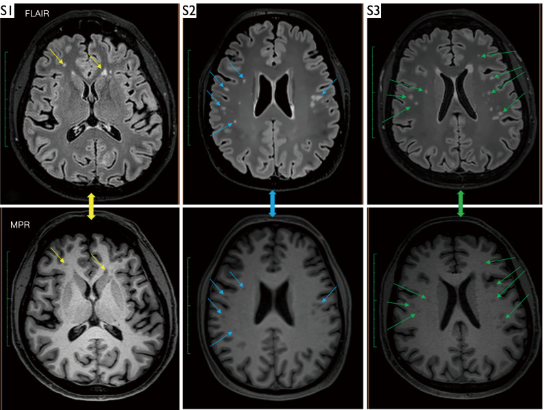
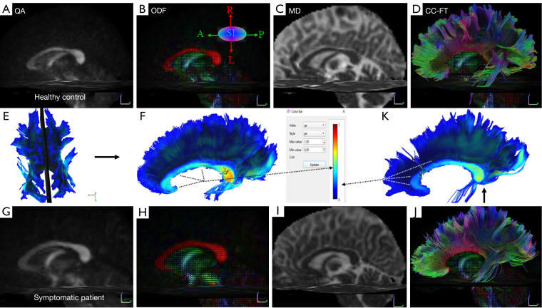
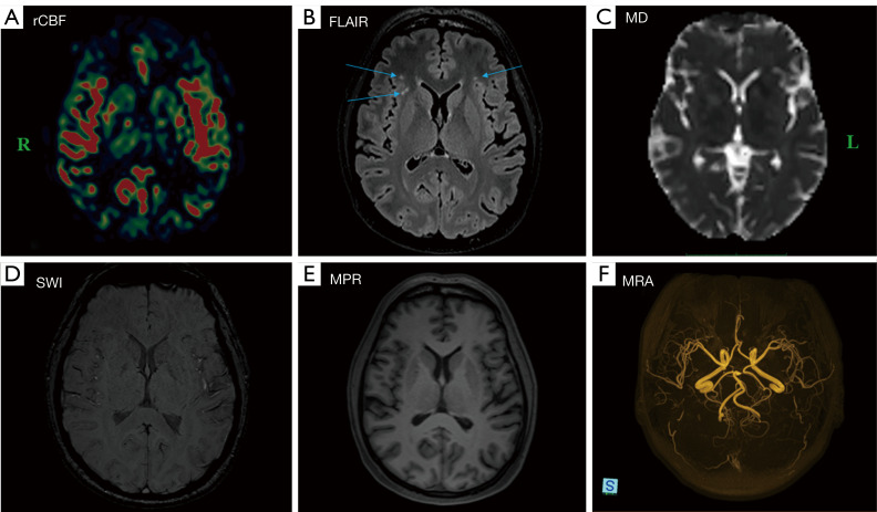
Statistical analysis revealed a significant difference in mean diffusivity (MD) between the symptomatic and control groups in the forceps minor (P=0.001) and CC body (P=0.003). In addition to changes in diffusion, alterations in brain perfusion were observed in two post-COVID-19 patients who experienced a severe course. Furthermore, hyperintense lesions were identified in subcortical and deep white matter areas in the vast majority of symptomatic patients.
The main finding of our study was that post-COVID-19 patients exhibit increased MD in the forceps minor and body of the CC. This finding suggests a potential association between microstructural brain changes in post-COVID-19 patients and reported neurological symptoms, with significant implications for research and clinical applications.
2019年冠状病毒病(COVID-19)先前发作后,血管系统和脑组织均可发生变化,可通过磁共振成像(MRI)技术检测扩散参数的改变来发现。这些扩散参数的变化在高度有组织的结构如胼胝体(CC)中可能尤为突出,包括其主要成分,而在COVID-19感染后尚未对其进行充分研究。因此,本研究旨在评估全脑(WB)扩散的微观结构变化,特别关注胼胝体。
共有101名受试者(年龄范围为18至69岁)参与了这项回顾性研究,其中包括55名志愿者和46名有神经症状的COVID-19康复患者。参与者于2022年4月至2023年9月在捷克共和国布拉格的临床与实验医学研究所招募。所有参与者均在3T MR扫描仪上进行了MRI检查,采用了扩散协议,并辅以其他MRI技术。由于运动伪影、严重灌注不足或存在病变,两名志愿者和五名患者被排除在研究之外。参与者由神经科医生根据临床检查和COVID-19抗体血清学检测进行选择。然后将他们分为三组:健康志愿者对照组(n=28)、有感染史但无症状的无症状组(n=25)和有COVID-19病史及神经症状的有症状组(n=41)。有症状的患者在感染COVID-19之前未表现出神经症状。扩散数据进行了涡流和敏感性失真校正,并在DSI studio中使用默认参数进行了纤维追踪。随后,在WB和CC的重建束内计算了各种扩散指标。为了评估COVID-19及其相关症状对WB和CC区域白质内扩散指数的影响,同时考虑年龄,我们在R框架内使用线性混合效应模型进行了统计分析。
统计分析显示,有症状组与对照组在小钳(P=0.001)和CC体(P=0.003)的平均扩散率(MD)上存在显著差异。除了扩散变化外,在两名经历严重病程的COVID-19康复患者中观察到脑灌注改变。此外,绝大多数有症状患者的皮质下和深部白质区域发现了高强度病变。
我们研究的主要发现是,COVID-19康复患者在小钳和CC体中表现出MD增加。这一发现表明COVID-19康复患者脑微观结构变化与报告的神经症状之间可能存在关联,对研究和临床应用具有重要意义。