Sakhawat Azra, Awan Sana Javaid, Khan Muhammad Umer, Shahid Samiah, Maqbool Tahir, Zubair Hafiz Muhammad, Manzoor Hina, Khan Samiullah
Institute of Molecular Biology and Biotechnology, The University of Lahore, Lahore, Pakistan.
Department of Biotechnology, Kinnaird College for Women University, Lahore, Pakistan.
J Taibah Univ Med Sci. 2024 Jul 10;19(4):775-789. doi: 10.1016/j.jtumed.2024.06.008. eCollection 2024 Aug.
This study was conducted to evaluate the effects of vitamin C on apoptotic and proliferative genes in injured HepG2 cells.
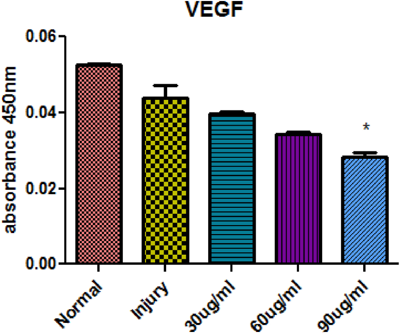
analysis was performed using molecular docking of chemical compounds with vascular endothelial growth factor (VEGF). The different computational tools used were AutoDock Vina, BIOVIA DISCOVERY studio, and PyMOL. Drug likeness and toxicity were analyzed by SWISS ADMET. Cells that were 60-70% confluent were treated with different concentrations of hydrogen peroxide (HO) (100-2000 μM) and ascorbic acid (30, 60, 90 μg/mL). The MTT cell proliferation assay was performed to compare the proliferative potential of HepG2 cells treated with HO or ascorbic acid with untreated HepG2 cells using 96-well plates.
The lowest binding energy of VEGF with vitamin C -5.2 kcal/mol and L-ascorbic acid-2 glycoside -4.7 kcal/mol was observed by analysis. Vitamin C was selected because it exhibited a high interaction with VEGF and fulfilled Lipinski's rule, and had better oral viability and pharmacokinetics compared to L-ascorbic acid-2 glycoside. Cell viability assays showed that vitamin C had significant apoptotic effects (P < 0.0001). After treating HepG2 cells with ascorbic acid, reduced VEGF (angiogenesis) was observed as determined by apoptotic and proliferative gene expression. Ascorbic acid treatment of HepG2 cells led to downregulation of the proliferation markers, proliferating cell nuclear antigen, Ki67, and DNA topoisomerase II alpha. Increased apoptosis after treatment with vitamin C was observed due to upregulation of p53 and annexin V.
The results of this study showed that vitamin C inhibited the growth of cancer cells, thus protecting HepG2 cells from oxidative stress. Vitamin C exhibited antiproliferative activity as observed and , as well as by the inhibited expression of genes involved in protein synthesis.
本研究旨在评估维生素C对受损HepG2细胞中凋亡和增殖基因的影响。
使用化合物与血管内皮生长因子(VEGF)的分子对接进行分析。所使用的不同计算工具包括AutoDock Vina、BIOVIA DISCOVERY studio和PyMOL。通过SWISS ADMET分析药物相似性和毒性。将60 - 70%汇合的细胞用不同浓度的过氧化氢(HO)(100 - 2000 μM)和抗坏血酸(30、60、90 μg/mL)处理。使用96孔板进行MTT细胞增殖试验,以比较用HO或抗坏血酸处理的HepG2细胞与未处理的HepG2细胞的增殖潜力。
通过分析观察到VEGF与维生素C的最低结合能为 -5.2 kcal/mol,与L - 抗坏血酸 -2 - 糖苷的最低结合能为 -4.7 kcal/mol。选择维生素C是因为它与VEGF表现出高相互作用且符合Lipinski规则,并且与L - 抗坏血酸 -2 - 糖苷相比具有更好的口服活性和药代动力学。细胞活力测定表明维生素C具有显著的凋亡作用(P < 0.0001)。用抗坏血酸处理HepG2细胞后,通过凋亡和增殖基因表达测定观察到VEGF(血管生成)减少。抗坏血酸处理HepG2细胞导致增殖标志物、增殖细胞核抗原、Ki67和DNA拓扑异构酶IIα的下调。由于p53和膜联蛋白V的上调,观察到用维生素C处理后凋亡增加。
本研究结果表明维生素C抑制癌细胞生长,从而保护HepG2细胞免受氧化应激。如分析和观察到的以及通过参与蛋白质合成的基因的抑制表达所示,维生素C表现出抗增殖活性。