• 文献检索
  • 文档翻译
  • 深度研究
  • 学术资讯
  • Suppr Zotero 插件Zotero 插件
  • 邀请有礼
  • 套餐&价格
  • 历史记录
应用&插件
Suppr Zotero 插件Zotero 插件浏览器插件Mac 客户端Windows 客户端微信小程序
定价
高级版会员购买积分包购买API积分包
服务
文献检索文档翻译深度研究API 文档MCP 服务
关于我们
关于 Suppr公司介绍联系我们用户协议隐私条款
关注我们

Suppr 超能文献

核心技术专利:CN118964589B侵权必究
粤ICP备2023148730 号-1Suppr @ 2026

文献检索

告别复杂PubMed语法,用中文像聊天一样搜索,搜遍4000万医学文献。AI智能推荐,让科研检索更轻松。

立即免费搜索

文件翻译

保留排版,准确专业,支持PDF/Word/PPT等文件格式,支持 12+语言互译。

免费翻译文档

深度研究

AI帮你快速写综述,25分钟生成高质量综述,智能提取关键信息,辅助科研写作。

立即免费体验

腹腔脓毒症时释放的细菌 ATP 可引发局部和全身炎症。

Released bacterial ATP shapes local and systemic inflammation during abdominal sepsis.

机构信息

Department of Visceral Surgery and Medicine, Inselspital, Bern University Hospital, University Hospital of Bern, Bern, Switzerland.

Department for BioMedical Research, Visceral Surgery and Medicine, University Hospital of Bern, Bern, Switzerland.

出版信息

Elife. 2024 Aug 20;13:RP96678. doi: 10.7554/eLife.96678.

DOI:10.7554/eLife.96678
PMID:39163101
原文链接:https://pmc.ncbi.nlm.nih.gov/articles/PMC11335348/
Abstract

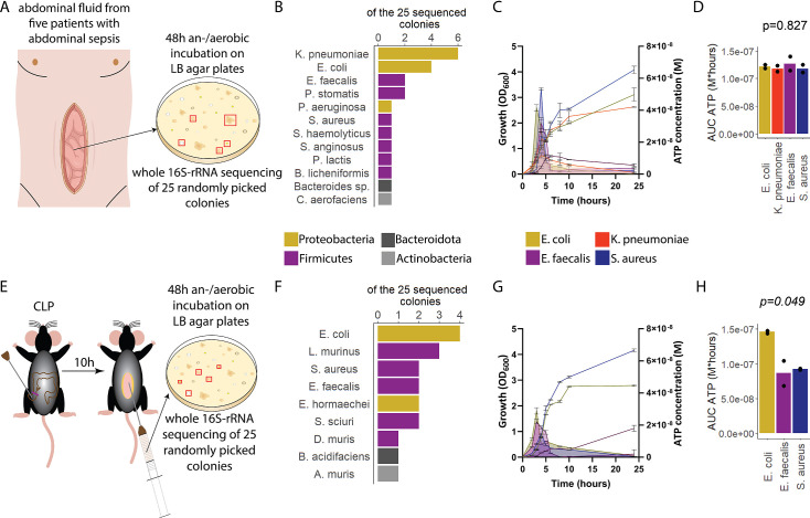
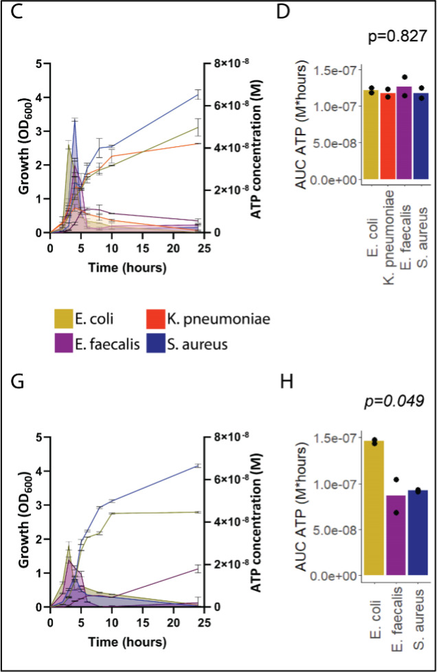
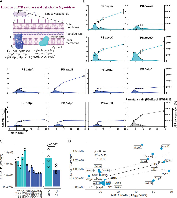
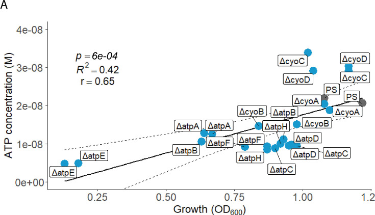
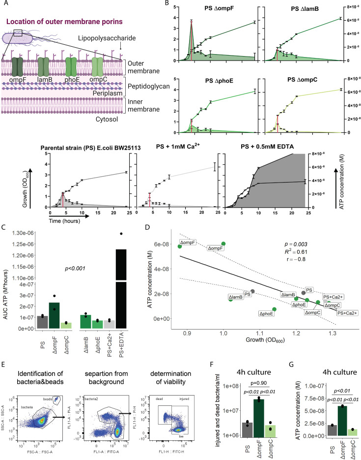
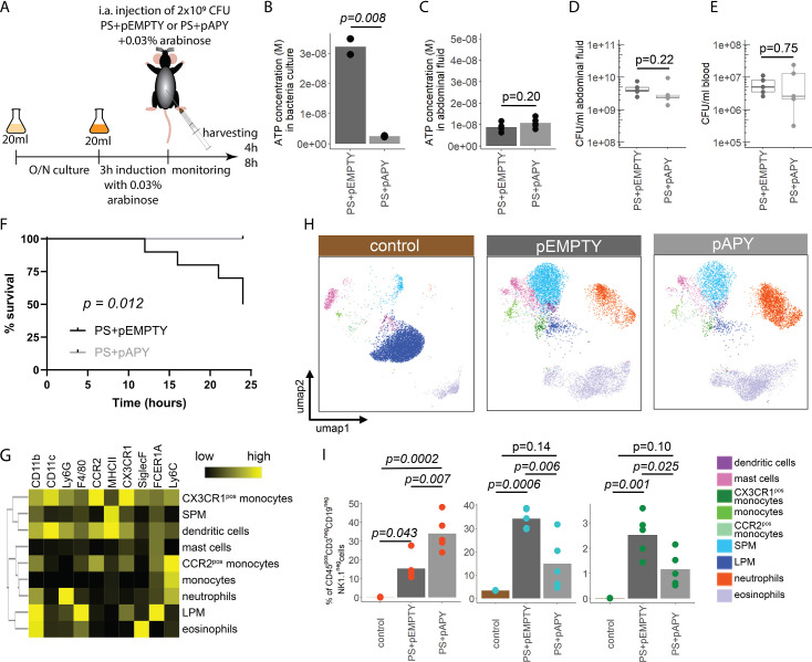
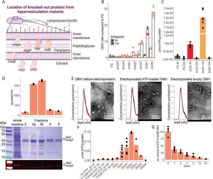
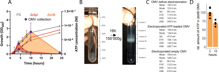
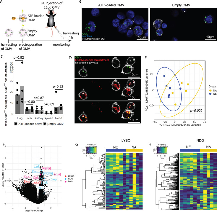
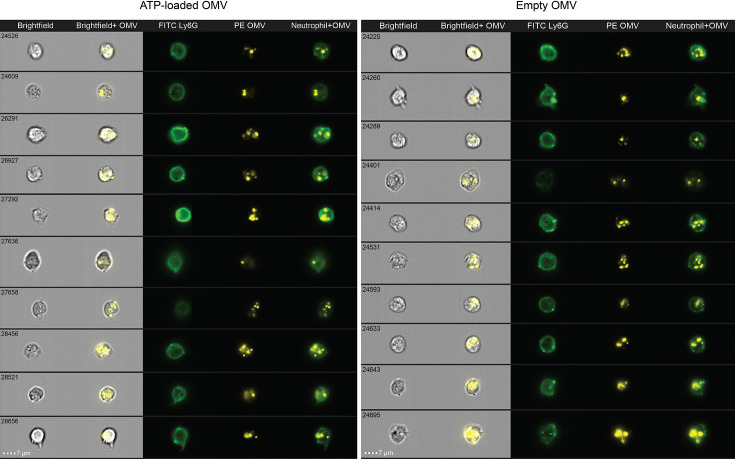
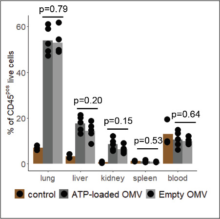
Sepsis causes millions of deaths per year worldwide and is a current global health priority declared by the WHO. Sepsis-related deaths are a result of dysregulated inflammatory immune responses indicating the need to develop strategies to target inflammation. An important mediator of inflammation is extracellular adenosine triphosphate (ATP) that is released by inflamed host cells and tissues, and also by bacteria in a strain-specific and growth-dependent manner. Here, we investigated the mechanisms by which bacteria release ATP. Using genetic mutant strains of (), we demonstrate that ATP release is dependent on ATP synthase within the inner bacterial membrane. In addition, impaired integrity of the outer bacterial membrane notably contributes to ATP release and is associated with bacterial death. In a mouse model of abdominal sepsis, local effects of bacterial ATP were analyzed using a transformed bearing an arabinose-inducible periplasmic apyrase hydrolyzing ATP to be released. Abrogating bacterial ATP release shows that bacterial ATP suppresses local immune responses, resulting in reduced neutrophil counts and impaired survival. In addition, bacterial ATP has systemic effects via its transport in outer membrane vesicles (OMV). ATP-loaded OMV are quickly distributed throughout the body and upregulated expression of genes activating degranulation in neutrophils, potentially contributing to the exacerbation of sepsis severity. This study reveals mechanisms of bacterial ATP release and its local and systemic roles in sepsis pathogenesis.

摘要

败血症每年导致全球数百万人死亡,是世卫组织宣布的当前全球卫生重点。与败血症相关的死亡是由于炎症免疫反应失调所致,这表明需要制定针对炎症的策略。炎症的一个重要介质是细胞外三磷酸腺苷 (ATP),它由炎症宿主细胞和组织释放,细菌也以菌株特异性和生长依赖性的方式释放。在这里,我们研究了细菌释放 ATP 的机制。使用 ( ) 的遗传突变株,我们证明 ATP 的释放依赖于内膜中的 ATP 合酶。此外,细菌外膜完整性的受损显著促进了 ATP 的释放,并与细菌死亡相关。在腹部败血症的小鼠模型中,使用携带阿拉伯糖诱导的周质质膜内的外切核酸酶水解释放的 ATP 的转化 来分析细菌 ATP 的局部作用。消除细菌 ATP 的释放表明细菌 ATP 抑制了局部免疫反应,导致中性粒细胞计数减少和存活率降低。此外,细菌 ATP 通过其在外膜囊泡 (OMV) 中的运输具有全身作用。负载 ATP 的 OMV 迅速分布在全身,并上调中性粒细胞脱颗粒的基因表达,可能导致败血症严重程度恶化。这项研究揭示了细菌 ATP 释放的机制及其在败血症发病机制中的局部和全身作用。

https://cdn.ncbi.nlm.nih.gov/pmc/blobs/c515/11335348/478ceb98af3a/elife-96678-sa3-fig2.jpg
https://cdn.ncbi.nlm.nih.gov/pmc/blobs/c515/11335348/68ed93c2e50d/elife-96678-fig1.jpg
https://cdn.ncbi.nlm.nih.gov/pmc/blobs/c515/11335348/57bb8be62c94/elife-96678-fig1-figsupp1.jpg
https://cdn.ncbi.nlm.nih.gov/pmc/blobs/c515/11335348/df2d82c55226/elife-96678-fig2.jpg
https://cdn.ncbi.nlm.nih.gov/pmc/blobs/c515/11335348/f78e28b079ab/elife-96678-fig2-figsupp1.jpg
https://cdn.ncbi.nlm.nih.gov/pmc/blobs/c515/11335348/52010d1bab1c/elife-96678-fig3.jpg
https://cdn.ncbi.nlm.nih.gov/pmc/blobs/c515/11335348/d42fd05700de/elife-96678-fig4.jpg
https://cdn.ncbi.nlm.nih.gov/pmc/blobs/c515/11335348/6c937e61da49/elife-96678-fig4-figsupp1.jpg
https://cdn.ncbi.nlm.nih.gov/pmc/blobs/c515/11335348/7e6b41368c25/elife-96678-fig5.jpg
https://cdn.ncbi.nlm.nih.gov/pmc/blobs/c515/11335348/72e4e6d00f1e/elife-96678-fig5-figsupp1.jpg
https://cdn.ncbi.nlm.nih.gov/pmc/blobs/c515/11335348/2560390c5513/elife-96678-fig6.jpg
https://cdn.ncbi.nlm.nih.gov/pmc/blobs/c515/11335348/28fe936385a0/elife-96678-fig6-figsupp1.jpg
https://cdn.ncbi.nlm.nih.gov/pmc/blobs/c515/11335348/26578747a89b/elife-96678-fig6-figsupp2.jpg
https://cdn.ncbi.nlm.nih.gov/pmc/blobs/c515/11335348/f53b8800031e/elife-96678-fig6-figsupp3.jpg
https://cdn.ncbi.nlm.nih.gov/pmc/blobs/c515/11335348/876e68a64763/elife-96678-fig6-figsupp4.jpg
https://cdn.ncbi.nlm.nih.gov/pmc/blobs/c515/11335348/f5cc0a2d0d5b/elife-96678-fig6-figsupp5.jpg
https://cdn.ncbi.nlm.nih.gov/pmc/blobs/c515/11335348/92cb716a3dce/elife-96678-sa3-fig1.jpg
https://cdn.ncbi.nlm.nih.gov/pmc/blobs/c515/11335348/478ceb98af3a/elife-96678-sa3-fig2.jpg

相似文献

1
Released bacterial ATP shapes local and systemic inflammation during abdominal sepsis.腹腔脓毒症时释放的细菌 ATP 可引发局部和全身炎症。
Elife. 2024 Aug 20;13:RP96678. doi: 10.7554/eLife.96678.
2
CD14 release induced by P2X7 receptor restricts inflammation and increases survival during sepsis.P2X7 受体诱导的 CD14 释放可限制脓毒症中的炎症反应并提高存活率。
Elife. 2020 Nov 2;9:e60849. doi: 10.7554/eLife.60849.
3
Frontline Science: Escherichia coli use LPS as decoy to impair neutrophil chemotaxis and defeat antimicrobial host defense.前沿科学:大肠杆菌利用脂多糖作为诱饵,削弱中性粒细胞趋化性并击败抗菌宿主防御。
J Leukoc Biol. 2019 Dec;106(6):1211-1219. doi: 10.1002/JLB.4HI0319-109R. Epub 2019 Aug 8.
4
Toll-like receptor-triggered calcium mobilization protects mice against bacterial infection through extracellular ATP release.Toll样受体触发的钙动员通过细胞外ATP释放保护小鼠免受细菌感染。
Infect Immun. 2014 Dec;82(12):5076-85. doi: 10.1128/IAI.02546-14. Epub 2014 Sep 22.
5
Immunization with Escherichia coli outer membrane vesicles protects bacteria-induced lethality via Th1 and Th17 cell responses.大肠杆菌外膜囊泡免疫通过 Th1 和 Th17 细胞应答保护细菌诱导的致死性。
J Immunol. 2013 Apr 15;190(8):4092-102. doi: 10.4049/jimmunol.1200742. Epub 2013 Mar 20.
6
A sustained type I IFN-neutrophil-IL-18 axis drives pathology during mucosal viral infection.持续的 I 型干扰素-中性粒细胞-白细胞介素 18 轴在黏膜病毒感染期间导致病理学改变。
Elife. 2021 May 28;10:e65762. doi: 10.7554/eLife.65762.
7
Colistin kills bacteria by targeting lipopolysaccharide in the cytoplasmic membrane.黏菌素通过靶向细胞质膜中的脂多糖杀死细菌。
Elife. 2021 Apr 6;10:e65836. doi: 10.7554/eLife.65836.
8
Systemic Adenosine Triphosphate Impairs Neutrophil Chemotaxis and Host Defense in Sepsis.全身性三磷酸腺苷损害脓毒症中性粒细胞趋化性及宿主防御功能。
Crit Care Med. 2017 Jan;45(1):e97-e104. doi: 10.1097/CCM.0000000000002052.
9
Decoding the complexity of delayed wound healing following infection.解析感染后伤口愈合延迟的复杂性。
Elife. 2024 May 20;13:RP95113. doi: 10.7554/eLife.95113.
10
Fructose-coated Ångstrom silver prevents sepsis by killing bacteria and attenuating bacterial toxin-induced injuries.果糖包裹的 Ångstrom 银通过杀死细菌和减轻细菌毒素引起的损伤来预防败血症。
Theranostics. 2021 Jul 13;11(17):8152-8171. doi: 10.7150/thno.55334. eCollection 2021.

引用本文的文献

1
The role of bacterial outer membrane vesicles in inflammatory response of acute-on-chronic liver failure.细菌外膜囊泡在慢加急性肝衰竭炎症反应中的作用
Front Microbiol. 2025 Jul 4;16:1608137. doi: 10.3389/fmicb.2025.1608137. eCollection 2025.
2
Outer membrane vesicles from Pseudomonas aeruginosa induce autophagy-regulated pyroptosis in THP-1 cells.铜绿假单胞菌的外膜囊泡可诱导THP-1细胞发生自噬调节的焦亡。
Arch Microbiol. 2025 Feb 10;207(3):54. doi: 10.1007/s00203-025-04264-9.
3
Horizontal gene transfer and beyond: the delivery of biological matter by bacterial membrane vesicles to host and bacterial cells.

本文引用的文献

1
Immune dysregulation in sepsis: experiences, lessons and perspectives.脓毒症中的免疫失调:经验、教训与展望。
Cell Death Discov. 2023 Dec 19;9(1):465. doi: 10.1038/s41420-023-01766-7.
2
Gata6 large peritoneal macrophages: an evolutionarily conserved sentinel and effector system for infection and injury.Gata6大型腹膜巨噬细胞:一种用于感染和损伤的进化保守的哨兵和效应系统。
Trends Immunol. 2023 Feb;44(2):129-145. doi: 10.1016/j.it.2022.12.002. Epub 2023 Jan 7.
3
Outer Membrane Vesicles: Biogenesis, Functions, and Issues.外膜囊泡:生物发生、功能和问题。
水平基因转移及其他:细菌膜泡向宿主细胞和细菌细胞递送生物物质
Curr Opin Microbiol. 2024 Oct;81:102525. doi: 10.1016/j.mib.2024.102525. Epub 2024 Aug 26.
Microbiol Mol Biol Rev. 2022 Dec 21;86(4):e0003222. doi: 10.1128/mmbr.00032-22. Epub 2022 Sep 26.
4
Outer membrane vesicles as molecular biomarkers for Gram-negative sepsis: Taking advantage of nature's perfect packages.外膜囊泡作为革兰氏阴性菌脓毒症的分子生物标志物:利用大自然完美的包装。
J Biol Chem. 2022 Oct;298(10):102483. doi: 10.1016/j.jbc.2022.102483. Epub 2022 Sep 13.
5
Optimised Electroporation for Loading of Extracellular Vesicles with Doxorubicin.用于用阿霉素加载细胞外囊泡的优化电穿孔法。
Pharmaceutics. 2021 Dec 24;14(1):38. doi: 10.3390/pharmaceutics14010038.
6
Use to Effectively Utilize Plotting Space to Deal With Large Datasets and Outliers.使用以有效利用绘图空间来处理大型数据集和异常值。
Front Genet. 2021 Nov 2;12:774846. doi: 10.3389/fgene.2021.774846. eCollection 2021.
7
Structure of Escherichia coli cytochrome bd-II type oxidase with bound aurachin D.结合金雀花碱 D 的大肠杆菌细胞色素 bd-II 型氧化酶的结构。
Nat Commun. 2021 Nov 11;12(1):6498. doi: 10.1038/s41467-021-26835-2.
8
Resident macrophage-dependent immune cell scaffolds drive anti-bacterial defense in the peritoneal cavity.驻留巨噬细胞依赖性免疫细胞支架驱动腹腔内的抗细菌防御。
Immunity. 2021 Nov 9;54(11):2578-2594.e5. doi: 10.1016/j.immuni.2021.10.007. Epub 2021 Oct 29.
9
Visualizing transfer of microbial biomolecules by outer membrane vesicles in microbe-host-communication in vivo.可视化外膜囊泡在微生物-宿主通讯中微生物生物分子转移。
J Extracell Vesicles. 2021 Oct;10(12):e12159. doi: 10.1002/jev2.12159.
10
The ATP Synthase Deficiency in Human Diseases.人类疾病中的ATP合酶缺乏症
Life (Basel). 2021 Apr 8;11(4):325. doi: 10.3390/life11040325.