Bitencourt Almir Galvão Vieira, Bhowmik Arka, Marcal Filho Eduardo Flavio De Lacerda, Lo Gullo Roberto, Mazaheri Yousef, Kapetas Panagiotis, Eskreis-Winkler Sarah, Young Robert, Pinker Katja, Thakur Sunitha B
Imaging Department, A. C. Camargo Cancer Center, São Paulo, 01525-001, Brazil.
Diagnósticos da América S.A., São Paulo, 04321-120, Brazil.
BJR Open. 2024 Aug 5;6(1):tzae019. doi: 10.1093/bjro/tzae019. eCollection 2024 Jan.
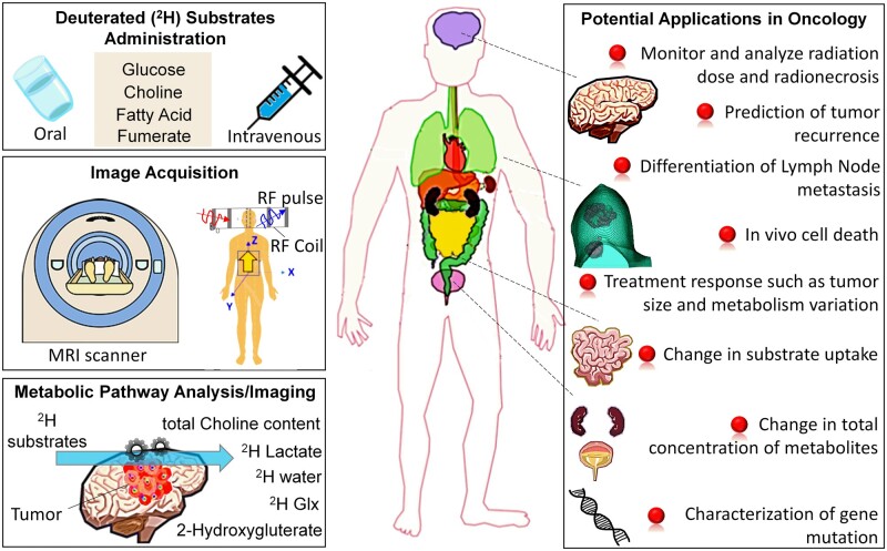
Metabolic imaging in clinical practice has long relied on PET with fluorodeoxyglucose (FDG), a radioactive tracer. However, this conventional method presents inherent limitations such as exposure to ionizing radiation and potential diagnostic uncertainties, particularly in organs with heightened glucose uptake like the brain. This review underscores the transformative potential of traditional deuterium MR spectroscopy (MRS) when integrated with gradient techniques, culminating in an advanced metabolic imaging modality known as deuterium MRI (DMRI). While recent advancements in hyperpolarized MRS hold promise for metabolic analysis, their widespread clinical usage is hindered by cost constraints and the availability of hyperpolarizer devices or facilities. DMRI, also denoted as deuterium metabolic imaging (DMI), represents a pioneering, single-shot, and noninvasive paradigm that fuses conventional MRS with nonradioactive deuterium-labelled substrates. Extensively tested in animal models and patient cohorts, particularly in cases of brain tumours, DMI's standout feature lies in its seamless integration into standard clinical MRI scanners, necessitating only minor adjustments such as radiofrequency coil tuning to the deuterium frequency. DMRI emerges as a versatile tool for quantifying crucial metabolites in clinical oncology, including glucose, lactate, glutamate, glutamine, and characterizing IDH mutations. Its potential applications in this domain are broad, spanning diagnostic profiling, treatment response monitoring, and the identification of novel therapeutic targets across diverse cancer subtypes.
临床实践中的代谢成像长期以来一直依赖于使用放射性示踪剂氟脱氧葡萄糖(FDG)的正电子发射断层扫描(PET)。然而,这种传统方法存在固有局限性,如暴露于电离辐射以及潜在的诊断不确定性,尤其是在葡萄糖摄取增加的器官如大脑中。本综述强调了传统的氘磁共振波谱(MRS)与梯度技术相结合时的变革潜力,最终形成了一种先进的代谢成像模式,即氘磁共振成像(DMRI)。虽然超极化MRS的最新进展为代谢分析带来了希望,但其广泛的临床应用受到成本限制以及超极化装置或设施可用性的阻碍。DMRI,也称为氘代谢成像(DMI),代表了一种开创性的、单次且无创的模式,它将传统的MRS与非放射性氘标记底物相结合。在动物模型和患者队列中进行了广泛测试,特别是在脑肿瘤病例中,DMI的突出特点在于它能够无缝集成到标准临床MRI扫描仪中,仅需进行诸如将射频线圈调谐到氘频率等微小调整。DMRI成为临床肿瘤学中量化关键代谢物(包括葡萄糖、乳酸、谷氨酸、谷氨酰胺)以及表征异柠檬酸脱氢酶(IDH)突变的通用工具。它在该领域的潜在应用广泛,涵盖诊断分析、治疗反应监测以及跨多种癌症亚型识别新的治疗靶点。