Biomedical MR Imaging and Spectroscopy Group, Center for Image Sciences, University Medical Center Utrecht and Utrecht University, Utrecht, the Netherlands.
Department of Radiology and Biomedical Imaging, Magnetic Resonance Research Center, Yale University School of Medicine, New Haven, CT, USA.
Neuroscience. 2021 Oct 15;474:94-99. doi: 10.1016/j.neuroscience.2021.01.023. Epub 2021 Jan 22.
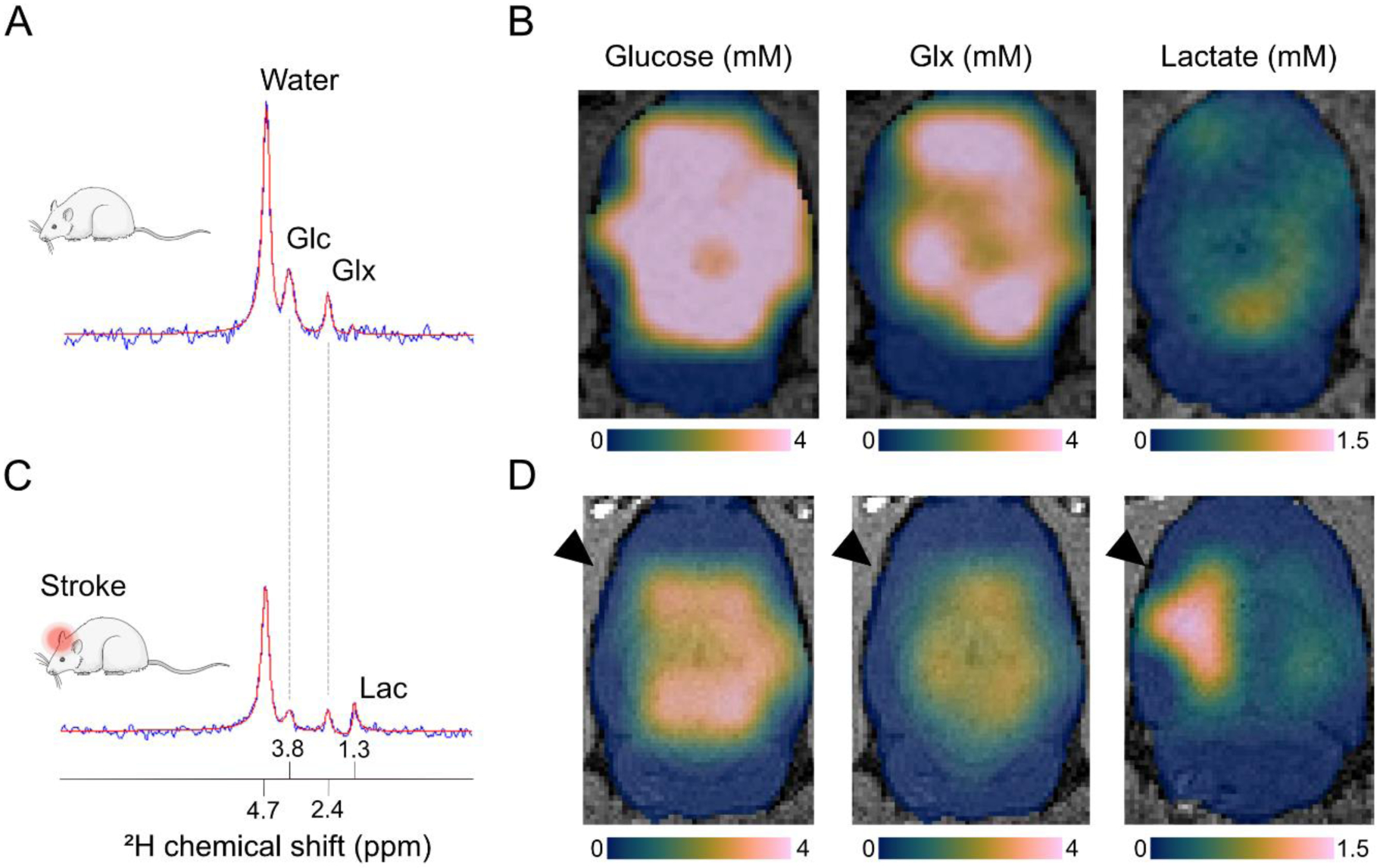
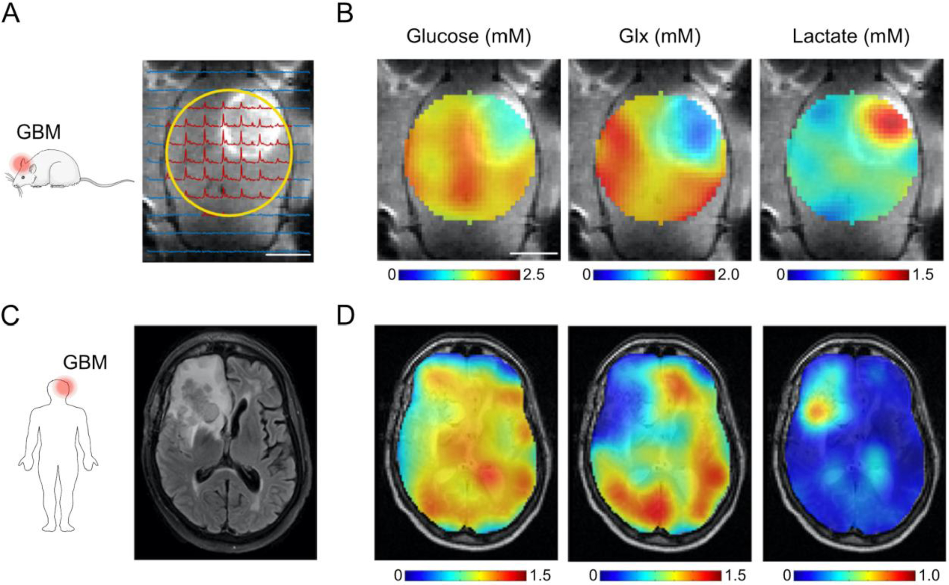
Altered brain metabolism contributes to pathophysiology in cerebrovascular and neurodegenerative diseases such as stroke and Alzheimer's disease. Current clinical tools to study brain metabolism rely on positron emission tomography (PET) requiring specific hardware and radiotracers, or magnetic resonance spectroscopy (MRS) involving technical complexity. In this review we highlight deuterium metabolic imaging (DMI) as a novel translational technique for assessment of brain metabolism, with examples from brain tumor and stroke studies. DMI is an MRS-based method that enables detection of deuterated substrates, such as glucose, and their metabolic products, such as lactate, glutamate and glutamine. It provides additional detail of downstream metabolites compared to analogous approaches like fluorodeoxyglucose (FDG)-PET, and can be implemented and executed on clinical and preclinical MR systems. We foresee that DMI, with future improvements in spatial and temporal resolutions, holds promise to become a valuable MR imaging (MRI) method for non-invasive mapping of glucose uptake and its downstream metabolites in healthy and diseased brain.
改变的大脑代谢有助于脑血管和神经退行性疾病的病理生理学,如中风和阿尔茨海默病。目前用于研究大脑代谢的临床工具依赖于正电子发射断层扫描(PET),需要特定的硬件和放射性示踪剂,或磁共振波谱(MRS),涉及技术复杂性。在这篇综述中,我们强调氘代谢成像(DMI)作为评估大脑代谢的一种新的转化技术,从脑肿瘤和中风研究中举例说明。DMI 是一种基于 MRS 的方法,可用于检测氘代底物,如葡萄糖,以及它们的代谢产物,如乳酸、谷氨酸和谷氨酰胺。与类似的方法(如氟代脱氧葡萄糖[FDG]-PET)相比,它提供了下游代谢物的更多细节,并且可以在临床和临床前磁共振系统上实现和执行。我们预计,随着空间和时间分辨率的未来提高,DMI 有望成为一种有价值的磁共振成像(MRI)方法,用于在健康和患病的大脑中无创性地映射葡萄糖摄取及其下游代谢物。