ANGLE Europe Limited, Guildford, UK.
Division of Gynecologic Oncology, Department of Obstetrics and Gynecology, Wilmot Cancer Institute, University of Rochester Medical Center, Rochester, NY, USA.
J Exp Clin Cancer Res. 2024 Aug 21;43(1):240. doi: 10.1186/s13046-024-03149-x.
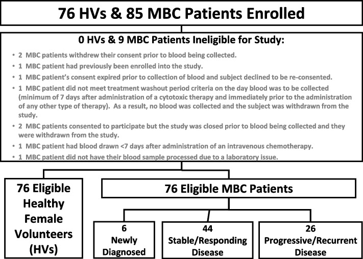
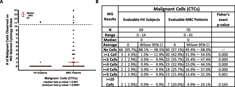
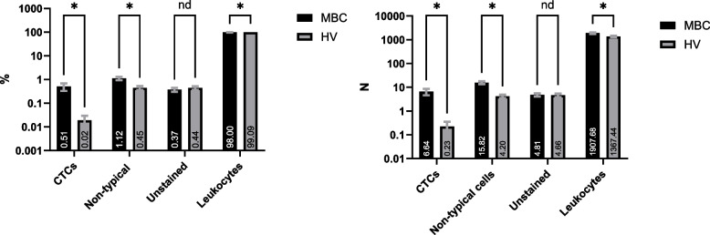
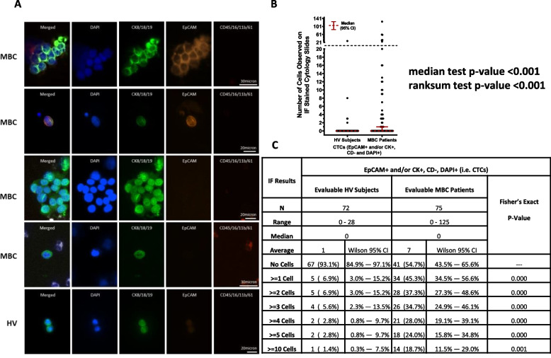
Circulating Tumor Cells (CTCs) may serve as a non-invasive source of tumor material to investigate an individual's disease in real-time. The Parsortix PC1 System, the first FDA-cleared medical device for the capture and harvest of CTCs from peripheral blood of metastatic breast cancer (MBC) patients for use in subsequent user-validated downstream analyses, enables the epitope-independent capture of CTCs with diverse phenotypes based on cell size and deformability. The aim of this study was to determine the proportion of MBC patients and self-declared female healthy volunteers (HVs) that had CTCs identified using immunofluorescence (IF) or Wright-Giemsa (WG) staining. Peripheral blood from 76 HVs and 76 MBC patients was processed on Parsortix PC1 Systems. Harvested cells were cytospun onto a charged slide and immunofluorescently stained for identification of CTCs expressing epithelial markers. The IF slides were subsequently WG-stained and analyzed for CTC identification based on morphological features of malignant cells. All testing was performed by operators blinded to the clinical status of each subject. CTCs were identified on the IF slides in 45.3% (≥ 1) / 24.0% (≥ 5) of the MBC patients (range = 0 - 125, mean = 7) and in 6.9% (≥ 1) / 2.8% (≥ 5) of the HVs (range = 0 - 28, mean = 1). Among the MBC patients with ≥ 1 CTC, 70.6% had only CK + /EpCAM- CTCs, with none having EpCAM + /CK- CTCs. CTC clusters were identified in 56.0% of the CTC-positive patients. On the WG-stained slides, CTCs were identified in 42.9% (≥ 1) / 21.4% (≥ 5) of the MBC patients (range = 0 - 41, mean = 4) and 4.3% (≥ 1) / 2.9% (≥ 5) of the HVs (range = 0 - 14, mean = 0). This study demonstrated the ability of the Parsortix PC1 System to capture and harvest CTCs from a significantly larger proportion of MBC patients compared to HVs when coupled with both IF and WG cytomorphological assessment. The presence of epithelial cells in subjects without diagnosed disease has been previously described, with their significance being unclear. Interestingly, a high proportion of the identified CTCs did not express EpCAM, highlighting the limitations of using EpCAM-based approaches.
循环肿瘤细胞(CTCs)可作为肿瘤物质的非侵入性来源,实时研究个体疾病。Parsortix PC1 系统是第一个获得 FDA 批准的用于捕获和收获转移性乳腺癌(MBC)患者外周血中 CTCs 的医疗器械,可用于随后经过用户验证的下游分析,能够基于细胞大小和变形性实现对具有不同表型的 CTCs 的表位独立捕获。本研究旨在确定使用免疫荧光(IF)或 Wright-Giemsa(WG)染色鉴定 CTCs 的 MBC 患者和自称女性健康志愿者(HV)的比例。对 76 名 HV 和 76 名 MBC 患者的外周血进行了 Parsortix PC1 系统处理。收获的细胞被细胞旋涂到带电荷的载玻片上,并通过免疫荧光染色鉴定表达上皮标志物的 CTC。随后对 IF 载玻片进行 WG 染色,并根据恶性细胞的形态特征分析 CTC 的鉴定。所有检测均由对每位受试者临床状况不知情的操作人员进行。在 45.3%(≥1)/24.0%(≥5)的 MBC 患者(范围=0-125,平均值=7)和 6.9%(≥1)/2.8%(≥5)的 HV 中(范围=0-28,平均值=1)在 IF 载玻片上鉴定出 CTC。在≥1 个 CTC 的 MBC 患者中,70.6%仅存在 CK+/EpCAM-CTC,没有 EpCAM+/CK-CTC。在 CTC 阳性患者中,CTC 簇的鉴定率为 56.0%。在 WG 染色载玻片上,在 42.9%(≥1)/21.4%(≥5)的 MBC 患者(范围=0-41,平均值=4)和 4.3%(≥1)/2.9%(≥5)的 HV 中(范围=0-14,平均值=0)鉴定出 CTC。本研究表明,与单独使用 IF 和 WG 细胞形态学评估相比,Parsortix PC1 系统能够从显着更多的 MBC 患者中捕获和收获 CTCs。在未诊断出疾病的受试者中存在上皮细胞先前已有描述,其意义尚不清楚。有趣的是,鉴定出的 CTC 中有很大一部分不表达 EpCAM,突出了使用基于 EpCAM 的方法的局限性。