Department of Gastro-Intestinal Surgery, University Hospital Ghent, Ghent, Belgium.
Center for Medical Genetics, Ghent University, Ghent, Belgium.
PLoS One. 2021 May 7;16(5):e0251052. doi: 10.1371/journal.pone.0251052. eCollection 2021.
The enrichment of circulating tumor cells (CTCs) from blood provides a minimally invasive method for biomarker discovery in cancer. Longitudinal interrogation allows monitoring or prediction of therapy response, detection of minimal residual disease or progression, and determination of prognosis. Despite inherent phenotypic heterogeneity and differences in cell surface marker expression, most CTC isolation technologies typically use positive selection. This necessitates the optimization of marker-independent CTC methods, enabling the capture of heterogenous CTCs. The aim of this report is to compare a size-dependent and a marker-dependent CTC-isolation method, using spiked esophageal cells in healthy donor blood and blood from patients diagnosed with esophageal adenocarcinoma.
Using esophageal cancer cell lines (OE19 and OE33) spiked into blood of a healthy donor, we investigated tumor cell isolation by Parsortix post cell fixation, immunostaining and transfer to a glass slide, and benchmarked its performance against the CellSearch system. Additionally, we performed DEPArray cell sorting to infer the feasibility to select and isolate cells of interest, aiming towards downstream single-cell molecular characterization in future studies. Finally, we measured CTC prevalence by Parsortix in venous blood samples from patients with various esophageal adenocarcinoma tumor stages.
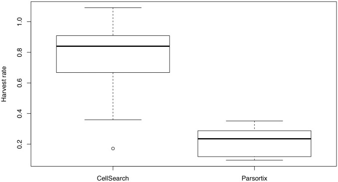
OE19 and OE33 cells were spiked in healthy donor blood and subsequently processed using CellSearch (n = 16) or Parsortix (n = 16). Upon tumor cell enrichment and enumeration, the recovery rate ranged from 76.3 ± 23.2% to 21.3 ± 9.2% for CellSearch and Parsortix, respectively. Parsortix-enriched and stained cell fractions were successfully transferred to the DEPArray instrument with preservation of cell morphology, allowing isolation of cells of interest. Finally, despite low CTC prevalence and abundance, Parsortix detected traditional CTCs (i.e. cytokeratin+/CD45-) in 8/29 (27.6%) of patients with esophageal adenocarcinoma, of whom 50% had early stage (I-II) disease.
We refined an epitope-independent isolation workflow to study CTCs in patients with esophageal adenocarcinoma. CTC recovery using Parsortix was substantially lower compared to CellSearch when focusing on the traditional CTC phenotype with CD45-negative and cytokeratin-positive staining characteristics. Future research could determine if this method allows downstream molecular interrogation of CTCs to infer new prognostic and predictive biomarkers on a single-cell level.
从血液中富集循环肿瘤细胞(CTC)为癌症的生物标志物发现提供了一种微创方法。纵向询问允许监测或预测治疗反应、检测微小残留疾病或进展,并确定预后。尽管存在固有表型异质性和细胞表面标志物表达的差异,但大多数 CTC 分离技术通常使用阳性选择。这需要优化与标记无关的 CTC 方法,从而实现异质 CTC 的捕获。本报告的目的是比较依赖大小和依赖标记的 CTC 分离方法,使用健康供体血液中的掺入食管细胞和诊断为食管腺癌的患者的血液。
使用掺入健康供体血液中的食管癌细胞系(OE19 和 OE33),我们研究了通过 Parsortix 后细胞固定、免疫染色和转移到载玻片上的肿瘤细胞分离,并将其性能与 CellSearch 系统进行了基准测试。此外,我们进行了 DEPArray 细胞分选,以推断选择和分离感兴趣细胞的可行性,旨在为未来的研究实现下游单细胞分子特征分析。最后,我们通过 Parsortix 测量了来自不同食管腺癌肿瘤阶段患者的静脉血样中的 CTC 患病率。
OE19 和 OE33 细胞被掺入健康供体血液中,然后使用 CellSearch(n=16)或 Parsortix(n=16)进行处理。在肿瘤细胞富集和计数后,CellSearch 和 Parsortix 的回收率分别为 76.3±23.2%至 21.3±9.2%。Parsortix 富集和染色的细胞分数成功转移到 DEPArray 仪器上,保留了细胞形态,允许分离感兴趣的细胞。最后,尽管 CTC 的患病率和丰度较低,但 Parsortix 在 29 名(27.6%)食管腺癌患者中检测到传统的 CTC(即角蛋白+/CD45-),其中 50%为早期(I-II)疾病。
我们改进了一种不依赖表位的分离工作流程,以研究食管腺癌患者的 CTC。与 CellSearch 相比,当重点关注具有 CD45 阴性和细胞角蛋白阳性染色特征的传统 CTC 表型时,Parsortix 用于 CTC 回收的效果要低得多。未来的研究可以确定该方法是否允许对 CTC 进行下游分子分析,以推断单细胞水平的新预后和预测生物标志物。