Genome Dynamics Laboratory, National Institute of Genetics, Mishima, Shizuoka 411-8540, Japan.
Graduate Institute for Advanced Studies (SOKENDAI), Mishima, Shizuoka 411-8540, Japan.
Proc Natl Acad Sci U S A. 2024 Sep 3;121(36):e2403153121. doi: 10.1073/pnas.2403153121. Epub 2024 Aug 27.
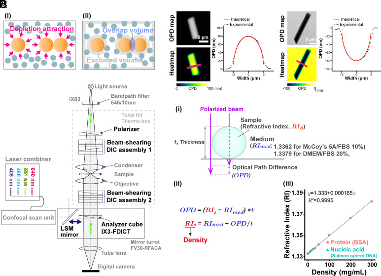
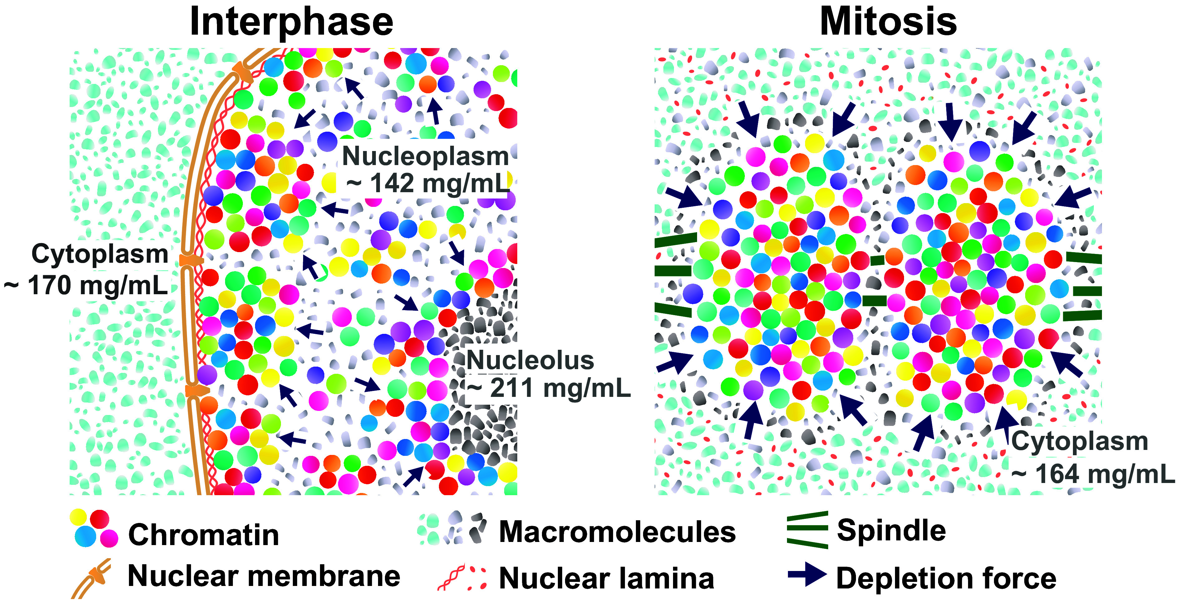
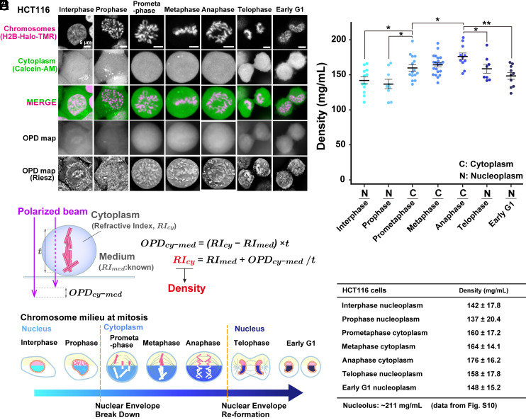
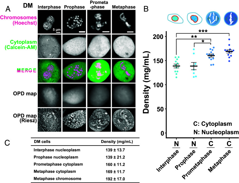
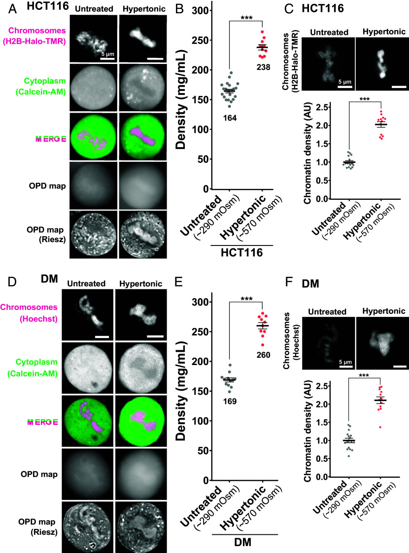
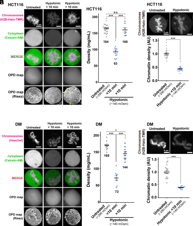
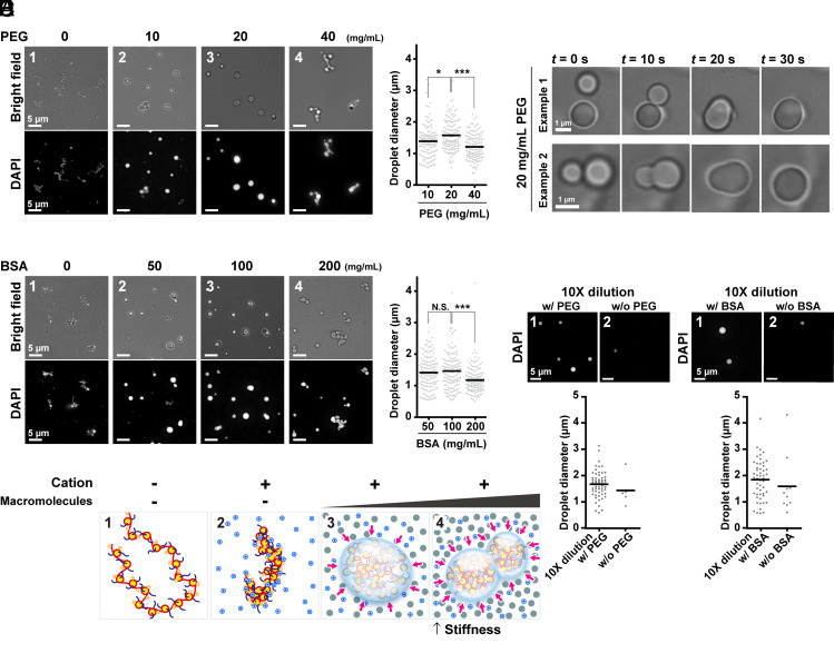
Genomic information must be faithfully transmitted into two daughter cells during mitosis. To ensure the transmission process, interphase chromatin is further condensed into mitotic chromosomes. Although protein factors like condensins and topoisomerase IIα are involved in the assembly of mitotic chromosomes, the physical bases of the condensation process remain unclear. Depletion attraction/macromolecular crowding, an effective attractive force that arises between large structures in crowded environments around chromosomes, may contribute to the condensation process. To approach this issue, we investigated the "chromosome milieu" during mitosis of living human cells using an orientation-independent-differential interference contrast module combined with a confocal laser scanning microscope, which is capable of precisely mapping optical path differences and estimating molecular densities. We found that the molecular density surrounding chromosomes increased with the progression from prophase to anaphase, concurring with chromosome condensation. However, the molecular density went down in telophase, when chromosome decondensation began. Changes in the molecular density around chromosomes by hypotonic or hypertonic treatment consistently altered the condensation levels of chromosomes. In vitro, native chromatin was converted into liquid droplets of chromatin in the presence of cations and a macromolecular crowder. Additional crowder made the chromatin droplets stiffer and more solid-like. These results suggest that a transient rise in depletion attraction, likely triggered by the relocation of macromolecules (proteins, RNAs, and others) via nuclear envelope breakdown and by a subsequent decrease in cell volumes, contributes to mitotic chromosome condensation, shedding light on a different aspect of the condensation mechanism in living human cells.
基因组信息必须在有丝分裂过程中忠实地传递到两个子细胞中。为了确保传递过程,间期染色质进一步浓缩成有丝分裂染色体。尽管像凝聚蛋白和拓扑异构酶 IIα 这样的蛋白质因子参与了有丝分裂染色体的组装,但浓缩过程的物理基础仍不清楚。耗散吸引/大分子拥挤,是在染色体周围拥挤环境中的大结构之间产生的一种有效吸引力,可能有助于浓缩过程。为了研究这个问题,我们使用一种不依赖于取向的差分干涉对比模块结合共聚焦激光扫描显微镜,对活的人类细胞有丝分裂过程中的“染色体环境”进行了研究,该显微镜能够精确地映射光路差异并估计分子密度。我们发现,随着从前期到后期的进展,染色体周围的分子密度增加,与染色体浓缩一致。然而,在末期,当染色体开始去浓缩时,分子密度下降。通过低渗或高渗处理改变染色体周围的分子密度,会一致地改变染色体的浓缩水平。在体外,在阳离子和大分子拥挤剂的存在下,天然染色质会转化为染色质的液滴。额外的拥挤剂使染色质液滴变得更硬,更类似固体。这些结果表明,耗散吸引的短暂上升,可能是由于核膜破裂导致大分子(蛋白质、RNA 等)的重定位,以及随后细胞体积的减少而触发的,有助于有丝分裂染色体的浓缩,为活的人类细胞中浓缩机制的不同方面提供了新的认识。