The Key Laboratory of Geriatrics, Beijing Institute of Geriatrics, Institute of Geriatric Medicine, Chinese Academy of Medical Sciences, Beijing Hospital/National Center of Gerontology of National Health Commission, NO.1 Da HuaRoad, DongDan, Beijing, 100730, People's Republic of China.
Cell Mol Biol Lett. 2024 Aug 28;29(1):113. doi: 10.1186/s11658-024-00623-4.
Cuproptosis is a unique copper-dependent form of cell death that is highly correlated with the metabolic state of cells. Triptolide exerts pharmacological activity by altering the regulation of metal ions. Cuproptosis is poorly understood in cancer, so in this study, we explored whether triptolide could induce cuproptosis in cervical cancer cells.
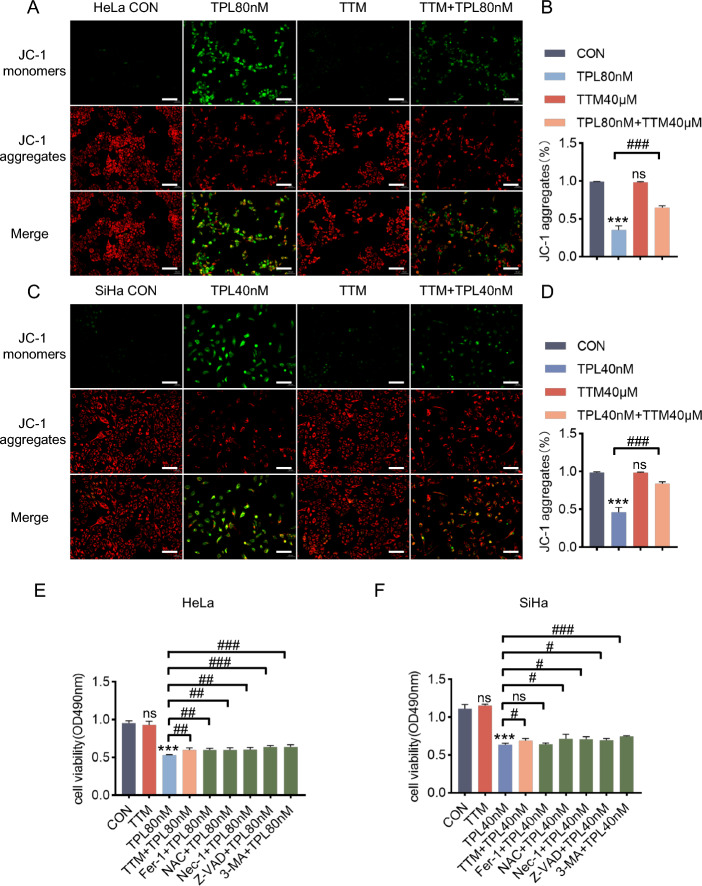
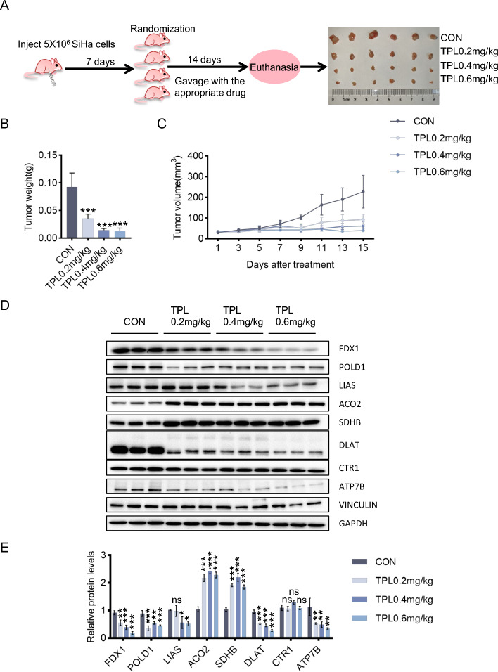
The human cervical cancer cell lines HeLa and SiHa, which primarily rely on oxidative phosphorylation, were treated with triptolide. Cell viability, proliferation and migration, copper levels and cuproptosis-related protein levels were evaluated in these cell lines. The copper ion chelator tetrathiomolybdate (TTM) was administered to determine whether it could reverse the cuproptosis induced by triptolide. In addition, a nude mouse cervical cancer xenograft model was established to determine the effects of triptolide on cuproptosis in isolated tumor tissues.
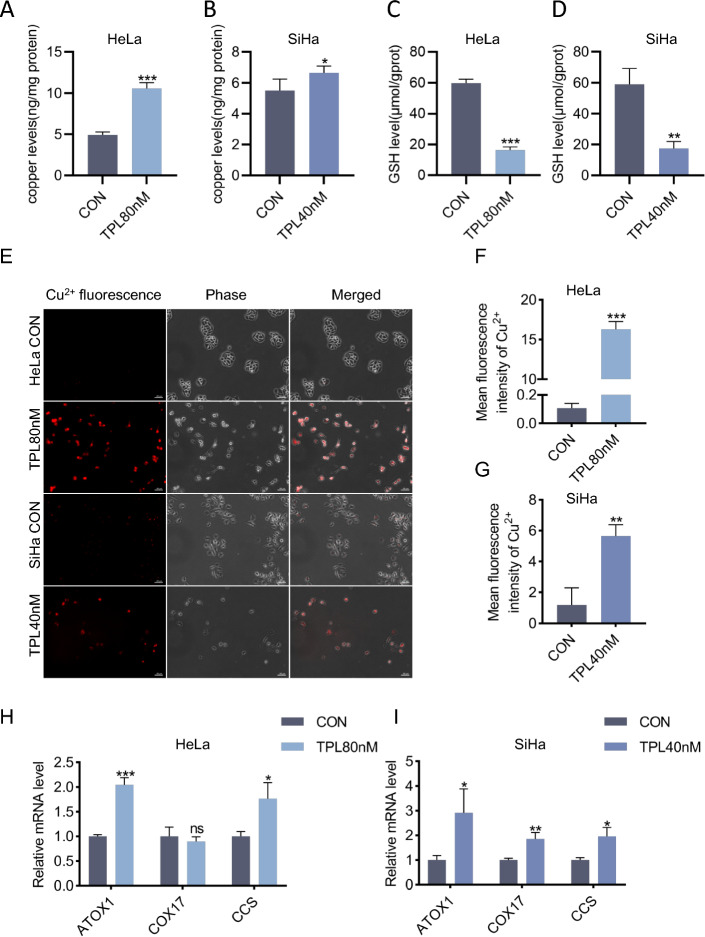
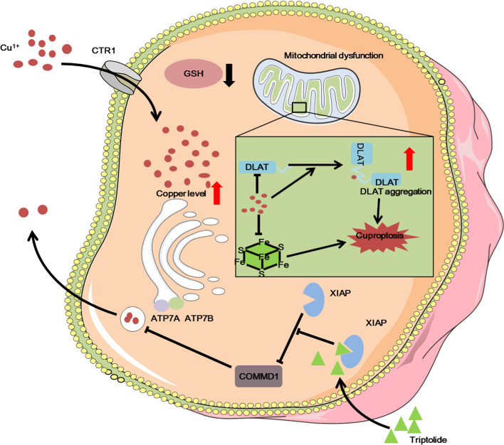
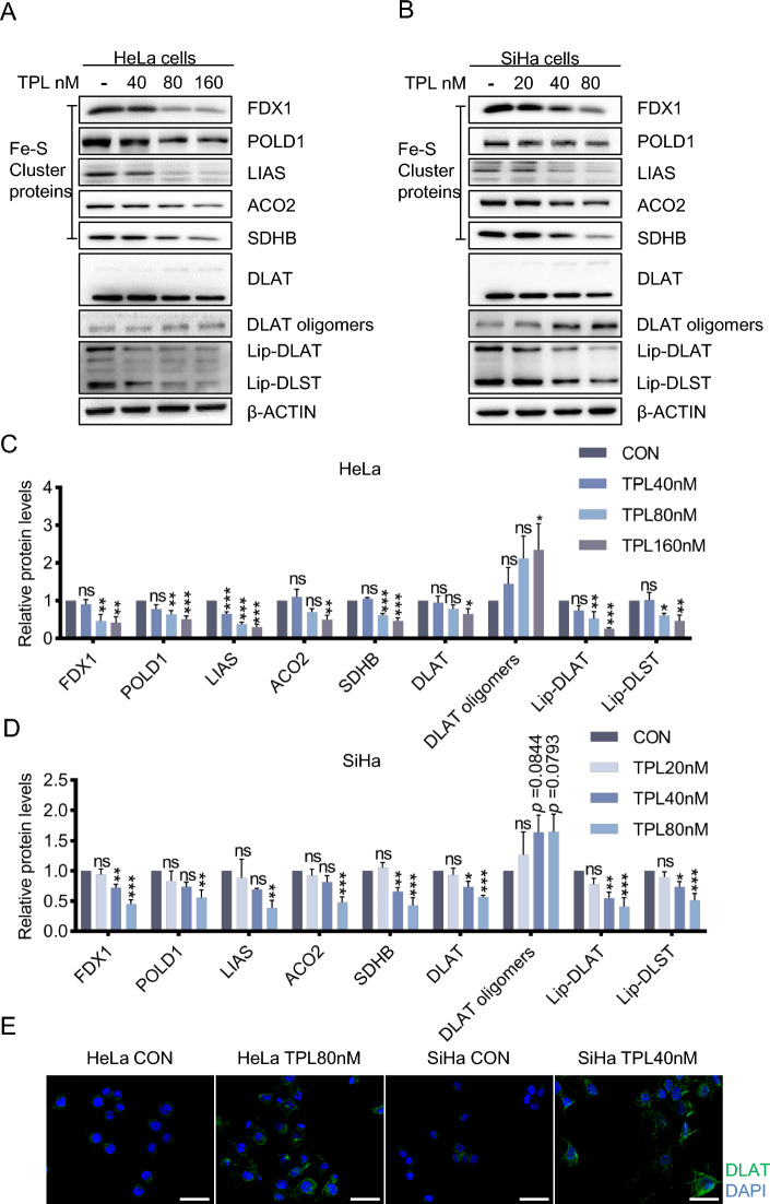
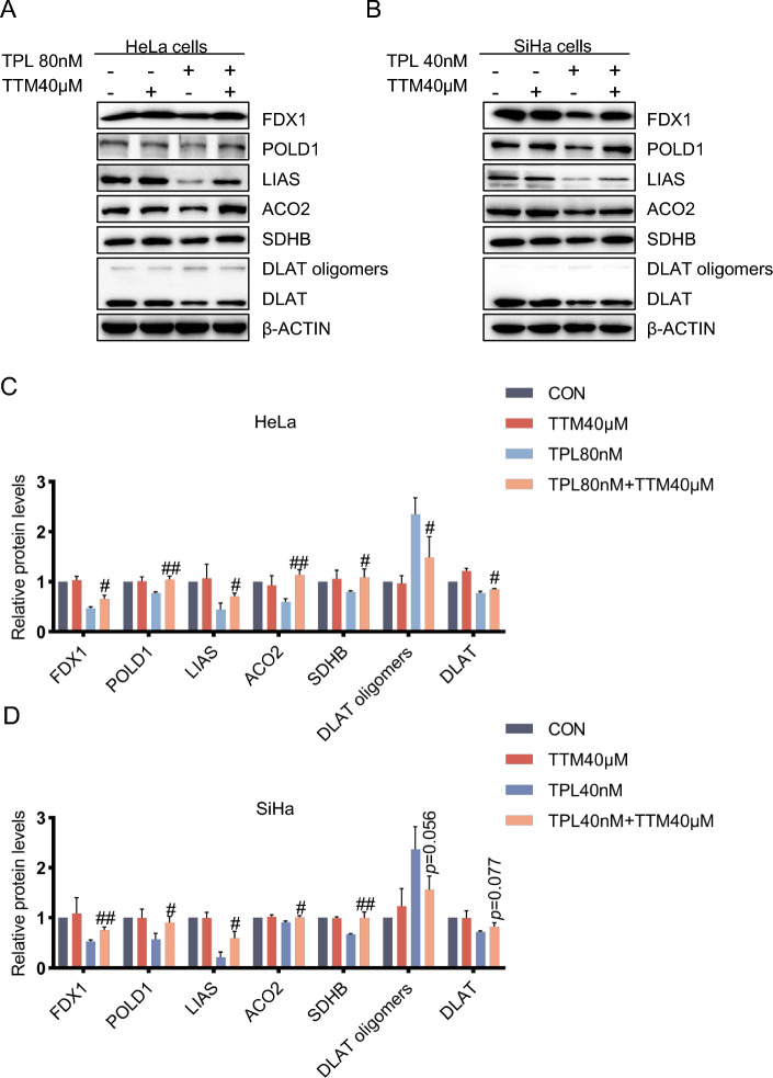
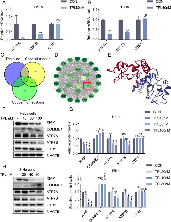
The copper concentration increased with triptolide treatment. The levels of cuproptosis -related proteins, such as FDX1, LIAS, and DLAT, in the HeLa and SiHa cell lines decreased with triptolide treatment. XIAP, the target of triptolide, played a role in cuproptosis by regulating COMMD1. The level of copper exporters (ATP7A/B) decreased, but the level of the copper importer (CTR1) did not change with triptolide treatment. Furthermore, triptolide inhibited cervical cancer growth and induced cuproptosis in vivo.
In summary, we report a new antitumor mechanism by which triptolide disrupted intracellular copper homeostasis and induced cuproptosis in cervical cancer by regulating the XIAP/COMMD1/ATP7A/B axis.
铜死亡是一种独特的、依赖铜的细胞死亡形式,与细胞的代谢状态高度相关。雷公藤红素通过改变金属离子的调节发挥药理活性。铜死亡在癌症中知之甚少,因此在这项研究中,我们探讨了雷公藤红素是否能诱导宫颈癌细胞发生铜死亡。
用雷公藤红素处理主要依赖氧化磷酸化的人宫颈癌细胞系 HeLa 和 SiHa。评估这些细胞系中的细胞活力、增殖和迁移、铜水平和铜死亡相关蛋白水平。给予铜离子螯合剂四硫钼酸盐(TTM),以确定其是否能逆转雷公藤红素诱导的铜死亡。此外,建立裸鼠宫颈癌异种移植模型,以确定雷公藤红素对分离的肿瘤组织中铜死亡的影响。
随着雷公藤红素处理,铜浓度增加。HeLa 和 SiHa 细胞系中与铜死亡相关的蛋白(如 FDX1、LIAS 和 DLAT)水平随着雷公藤红素处理而降低。雷公藤红素的靶蛋白 XIAP 通过调节 COMMD1 发挥铜死亡作用。铜外排蛋白(ATP7A/B)的水平降低,但铜内流蛋白(CTR1)的水平在雷公藤红素处理后没有变化。此外,雷公藤红素抑制宫颈癌的生长并在体内诱导铜死亡。
总之,我们报告了一种新的抗肿瘤机制,即雷公藤红素通过调节 XIAP/COMMD1/ATP7A/B 轴破坏细胞内铜平衡并诱导宫颈癌发生铜死亡。