Department of Surgery, School of Medicine, University of Mostar, University Hospital Mostar, Bijeli Brijeg bb, 88000 Mostar, Bosnia and Herzegovina.
Laboratory for Early Human Development, Department of Anatomy, Histology and Embryology, University of Split School of Medicine, Šoltanska 2A, 21000 Split, Croatia.
Medicina (Kaunas). 2024 Jul 25;60(8):1202. doi: 10.3390/medicina60081202.
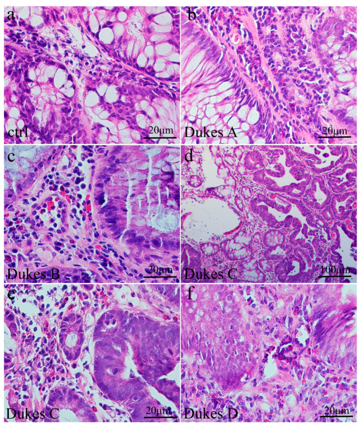
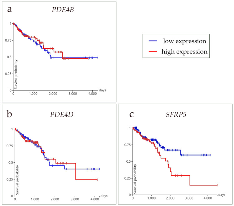
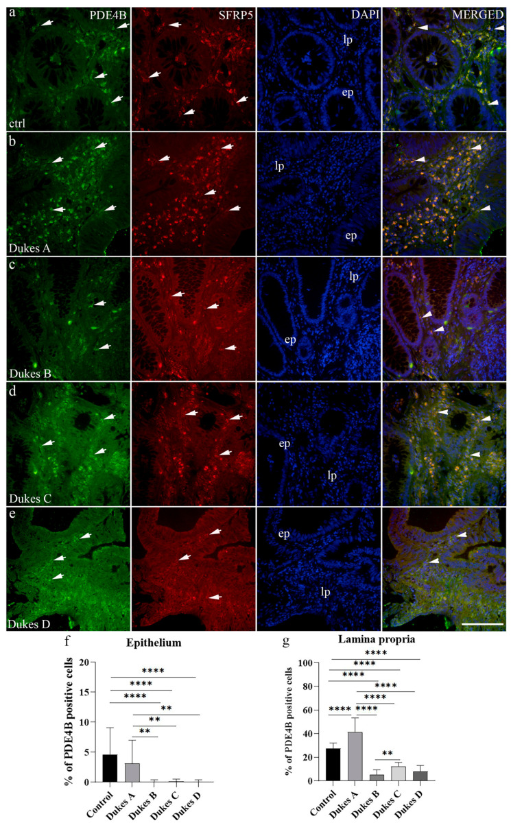
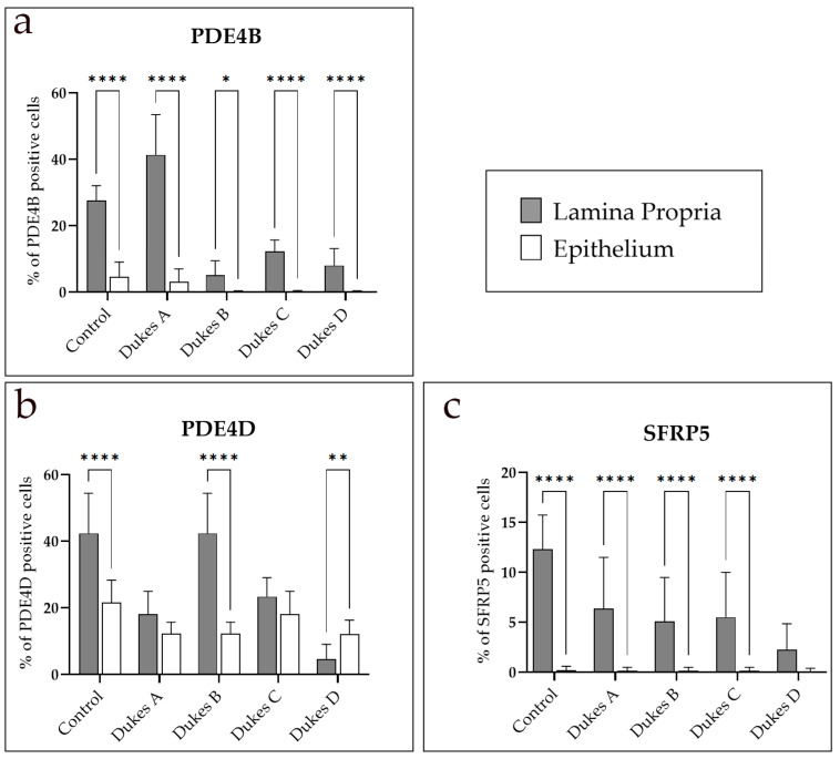
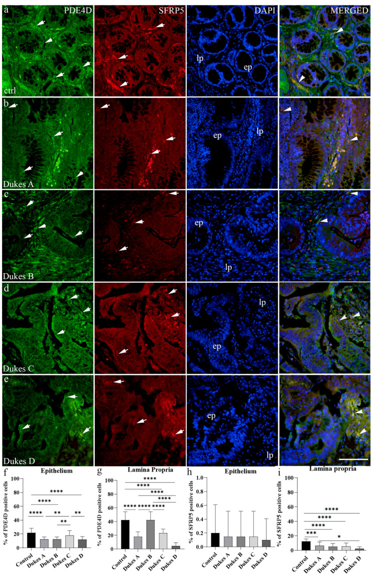
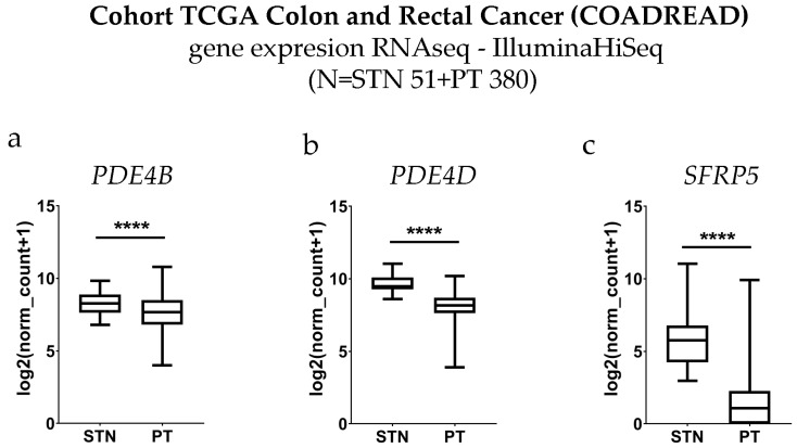
Colorectal cancer (CRC) is the most frequently diagnosed malignant disease of the gastrointestinal system, and new diagnostic and prognostic markers are needed to elucidate the complete tumor profile. : We used CRC tumor tissues (Dukes' A-D) and adjacent noncancerous tissues of 43 patients. Immunohistochemistry was used to examine the expression of phosphodiesterase 4B (PDE4B), phosphodiesterase 4D (PDE4D), and secreted frizzled related protein 5 (SFRP5) markers. We also analyzed the expression levels of , , and in CRC tissues compared to control tissues using RNA-sequencing data from the UCSC Xena browser. : In CRC stages, the distribution of PDE4B-positive cells varied, with differing percentages between epithelium and lamina propria. Statistically significant differences were found in the number of PDE4B-positive epithelial cells between healthy controls and all CRC stages, as well as between different CRC stages. Similarly, significant differences were observed in the number of PDE4B-positive cells in the lamina propria between healthy controls and all CRC stages, as well as between different CRC stages. CRC stage Dukes' C exhibited a significantly higher number of PDE4B-positive cells in the lamina propria compared to CRC stage Dukes' B. Significant differences were noted in the number of PDE4D-positive epithelial cells between healthy controls and CRC stages Dukes' A, B, and D, as well as between CRC stage Dukes' C and stages A, B, and D. CRC stage Dukes' A had significantly more PDE4D-positive cells in the lamina propria compared to stage D. Significant differences were also observed in the number of SFRP5-positive cells in the lamina propria between healthy controls and all CRC stages, as well as between CRC stages Dukes' A and D. While the expression of PDE4D varied across CRC stages, the expression of SFRP5 remained consistently strong in both epithelium and lamina propria, with significant differences noted mainly in the lamina propria. The expression levels of , , and reveal significant differences in the expression of these genes between CRC patients and healthy controls, with notable implications for patient prognosis. Namely, our results demonstrate that and are significantly under-expressed in CRC tissues compared to control tissues. The Kaplan-Meier survival analysis and the log-rank (Mantel-Cox) test revealed distinct prognostic implications where patients with lower expression levels of exhibited significantly longer overall survival. The data align with our immunohistochemical results and might suggest a potential tumor-suppressive role for these genes in CRC. : Considering significantly lower gene expression, aligned with our immunohistochemical data in tumor tissue in comparison to the control tissue, as well as the significantly poorer survival rate in the case of its higher expression, we can hypothesize that SFRP5 is the most promising biomarker for CRC out of the observed proteins. These findings suggest alterations in PDE4B, PDE4D, and SFRP5 expression during CRC progression, as well as between different stages of CRC, with potential implications for understanding the molecular mechanisms involved in CRC development and progression.
结直肠癌(CRC)是最常见的胃肠道系统恶性疾病,需要新的诊断和预后标志物来阐明完整的肿瘤特征。我们使用了 43 名患者的 CRC 肿瘤组织(Dukes'A-D)和相邻的非癌组织。采用免疫组织化学方法检测磷酸二酯酶 4B(PDE4B)、磷酸二酯酶 4D(PDE4D)和分泌卷曲相关蛋白 5(SFRP5)标志物的表达。我们还使用 UCSC Xena 浏览器中的 RNA 测序数据比较了 CRC 组织中与对照组织中 、 和 的表达水平。在 CRC 阶段,PDE4B 阳性细胞的分布不同,上皮和固有层之间的百分比不同。在健康对照组和所有 CRC 阶段以及不同 CRC 阶段之间,PDE4B 阳性上皮细胞的数量存在统计学显著差异。同样,在健康对照组和所有 CRC 阶段以及不同 CRC 阶段之间,PDE4B 阳性固有层细胞的数量也存在显著差异。CRC 阶段 Dukes'C 的固有层中 PDE4B 阳性细胞的数量明显高于 CRC 阶段 Dukes'B。健康对照组和 CRC 阶段 Dukes'A、B 和 D 之间以及 CRC 阶段 Dukes'C 和 A、B 和 D 之间,上皮细胞中 PDE4D 阳性细胞的数量存在显著差异。CRC 阶段 Dukes'A 的固有层中 PDE4D 阳性细胞明显多于阶段 D。固有层中 SFRP5 阳性细胞数量也存在显著差异,健康对照组和所有 CRC 阶段以及 CRC 阶段 Dukes'A 和 D 之间。虽然 PDE4D 的表达在 CRC 阶段有所不同,但 SFRP5 的表达在整个上皮和固有层中始终保持强烈,主要在上皮层中观察到显著差异。、 和 的表达水平显示这些基因在 CRC 患者和健康对照组之间的表达存在显著差异,这对患者的预后有重要意义。我们的结果表明,与对照组织相比,CRC 组织中 和 的表达显著下调。Kaplan-Meier 生存分析和对数秩(Mantel-Cox)检验显示出不同的预后意义,其中表达水平较低的患者总生存率显著延长。这些数据与我们的免疫组织化学结果一致,可能表明这些基因在 CRC 中具有潜在的肿瘤抑制作用。考虑到与对照组织相比,肿瘤组织中的基因表达明显下调,以及表达较高时的生存率明显较差,我们可以假设 SFRP5 是观察到的蛋白中最有前途的 CRC 生物标志物。这些发现表明,在 CRC 进展过程中以及在不同的 CRC 阶段,PDE4B、PDE4D 和 SFRP5 的表达发生改变,这可能有助于理解涉及 CRC 发生和进展的分子机制。