Department of Neurosciences, NC30, Lerner Research Institute, Cleveland Clinic, 9500 Euclid Avenue, Cleveland, OH, 44195, USA.
Department of Biomedical Engineering, Lerner Research Institute, Cleveland Clinic, Cleveland, OH, USA.
Acta Neuropathol. 2024 Aug 31;148(1):34. doi: 10.1007/s00401-024-02796-w.
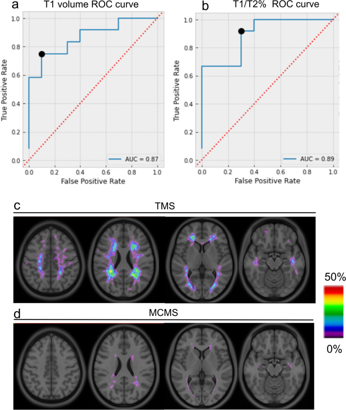
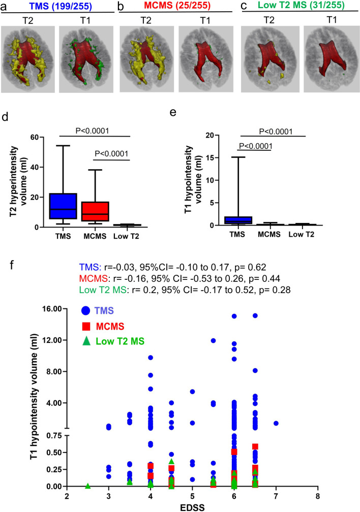
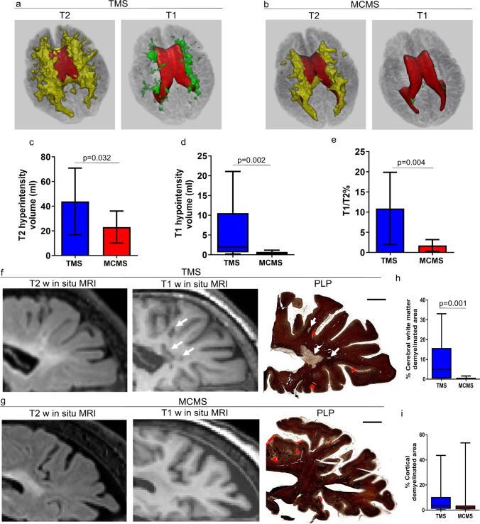
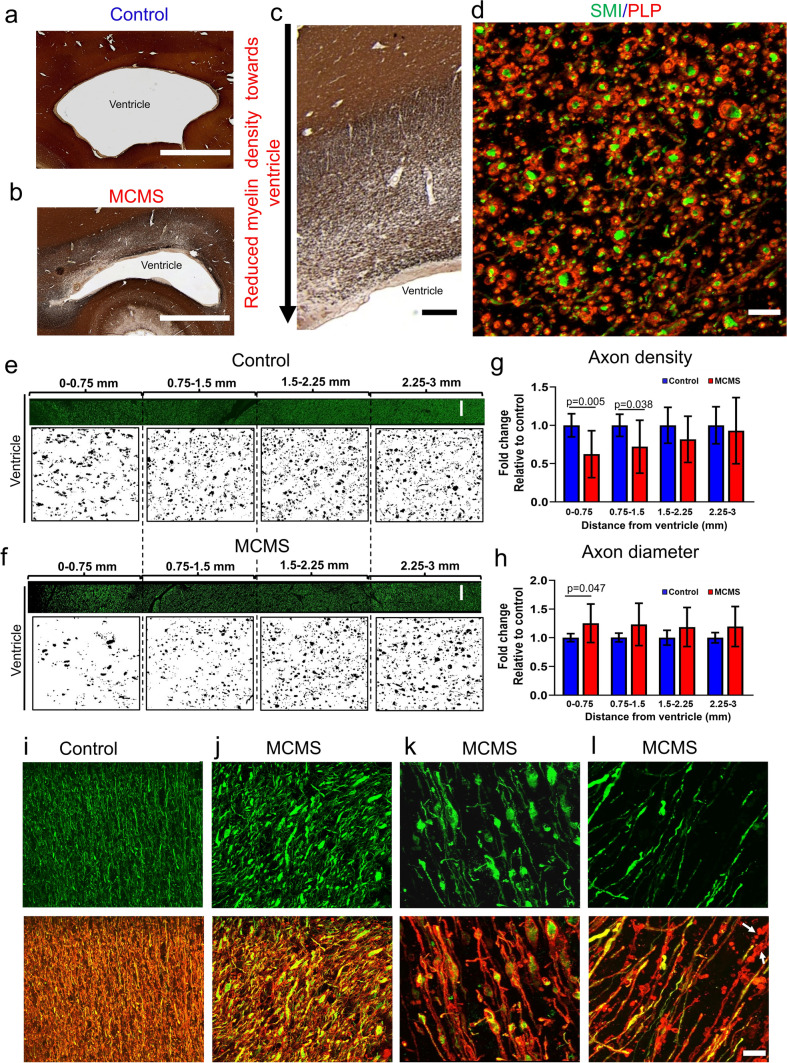
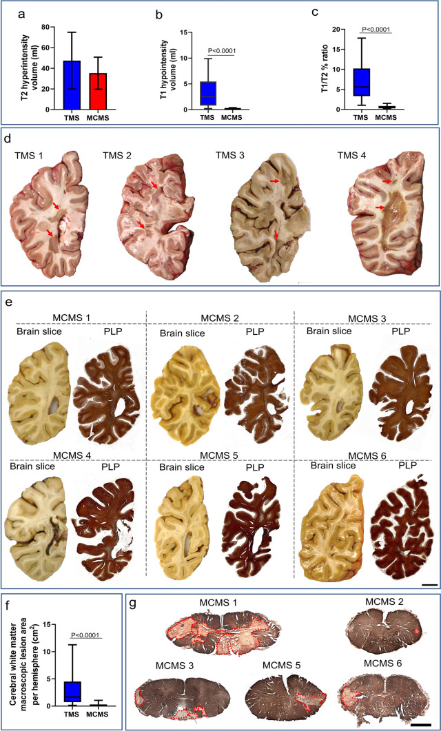
The pathogenic mechanisms contributing to neurological disability in progressive multiple sclerosis (PMS) are poorly understood. Cortical neuronal loss independent of cerebral white matter (WM) demyelination in myelocortical MS (MCMS) and identification of MS patients with widespread cortical atrophy and disability progression independent of relapse activity (PIRA) support pathogenic mechanisms other than cerebral WM demyelination. The three-dimensional distribution and underlying pathology of myelinated T2 lesions were investigated in postmortem MCMS brains. Postmortem brain slices from previously characterized MCMS (10 cases) and typical MS (TMS) cases (12 cases) were co-registered with in situ postmortem T2 hyperintensities and T1 hypointensities. T1 intensity thresholds were used to establish a classifier that differentiates MCMS from TMS. The classifier was validated in 36 uncharacterized postmortem brains and applied to baseline MRIs from 255 living PMS participants enrolled in SPRINT-MS. Myelinated T2 hyperintensities in postmortem MCMS brains have a contiguous periventricular distribution that expands at the occipital poles of the lateral ventricles where a surface-in gradient of myelinated axonal degeneration was observed. The MRI classifier distinguished pathologically confirmed postmortem MCMS and TMS cases with an accuracy of 94%. For SPRINT-MS patients, the MRI classifier identified 78% as TMS, 10% as MCMS, and 12% with a paucity of cerebral T1 and T2 intensities. In SPRINT-MS, expanded disability status scale and brain atrophy measures were similar in MCMS and TMS cohorts. A paucity of cerebral WM demyelination in 22% of living PMS patients raises questions regarding a primary role for cerebral WM demyelination in disability progression in all MS patients and has implications for clinical management of MS patients and clinical trial outcomes in PMS. Periventricular myelinated fiber degeneration provides additional support for surface-in gradients of neurodegeneration in MS.
导致进行性多发性硬化症(PMS)神经功能障碍的发病机制尚不清楚。在皮质脊髓 MS(MCMS)中,皮质神经元丢失与脑白质(WM)脱髓鞘无关,以及发现广泛皮质萎缩和残疾进展与复发活动无关的 MS 患者(PIRA),支持除 WM 脱髓鞘以外的发病机制。研究了死后 MCMS 大脑中髓鞘化 T2 病变的三维分布和潜在病理学。将以前特征明确的 MCMS(10 例)和典型 MS(TMS)病例(12 例)的死后脑切片与死后 T2 高信号和 T1 低信号的原位进行共配准。使用 T1 强度阈值建立一个分类器,将 MCMS 与 TMS 区分开来。该分类器在 36 例未经特征描述的死后大脑中进行了验证,并应用于 255 名参加 SPRINT-MS 的 PMS 活存患者的基线 MRI。死后 MCMS 大脑中的髓鞘化 T2 高信号具有连续的脑室周围分布,在侧脑室枕极处扩展,在该处观察到髓鞘化轴突变性的表面梯度。MRI 分类器对经病理证实的死后 MCMS 和 TMS 病例的区分准确率为 94%。对于 SPRINT-MS 患者,MRI 分类器将 78%识别为 TMS,10%为 MCMS,12%为脑 T1 和 T2 强度不足。在 SPRINT-MS 中,MCMS 和 TMS 队列的扩展残疾状态量表和脑萎缩测量值相似。22%的活存 PMS 患者脑 WM 脱髓鞘不足,这引发了关于 WM 脱髓鞘在所有 MS 患者残疾进展中起主要作用的问题,并对 MS 患者的临床管理和 PMS 临床试验结果产生影响。脑室周围髓鞘化纤维变性为 MS 中的神经退行性表面梯度提供了额外支持。