• 文献检索
  • 文档翻译
  • 深度研究
  • 学术资讯
  • Suppr Zotero 插件Zotero 插件
  • 邀请有礼
  • 套餐&价格
  • 历史记录
应用&插件
Suppr Zotero 插件Zotero 插件浏览器插件Mac 客户端Windows 客户端微信小程序
定价
高级版会员购买积分包购买API积分包
服务
文献检索文档翻译深度研究API 文档MCP 服务
关于我们
关于 Suppr公司介绍联系我们用户协议隐私条款
关注我们

Suppr 超能文献

核心技术专利:CN118964589B侵权必究
粤ICP备2023148730 号-1Suppr @ 2026

文献检索

告别复杂PubMed语法,用中文像聊天一样搜索,搜遍4000万医学文献。AI智能推荐,让科研检索更轻松。

立即免费搜索

文件翻译

保留排版,准确专业,支持PDF/Word/PPT等文件格式,支持 12+语言互译。

免费翻译文档

深度研究

AI帮你快速写综述,25分钟生成高质量综述,智能提取关键信息,辅助科研写作。

立即免费体验

耳蜗临床CT扫描的自动分割及耳蜗垂直轮廓分析。

Automated segmentation of clinical CT scans of the cochlea and analysis of the cochlea's vertical profile.

作者信息

Siebrecht Michael, Briaire Jeroen J, Verbist Berit M, Kalkman Randy K, Frijns Johan H M

机构信息

Department of Otorhinolaryngology and Head & Neck Surgery, Leiden University Medical Center, PO Box 9600, 2300 RC, Leiden, the Netherlands.

Leiden Institute for Brain and Cognition, PO Box 9600, 2300 RC, Leiden, the Netherlands.

出版信息

Heliyon. 2024 Aug 6;10(16):e35737. doi: 10.1016/j.heliyon.2024.e35737. eCollection 2024 Aug 30.

DOI:10.1016/j.heliyon.2024.e35737
PMID:39224385
原文链接:https://pmc.ncbi.nlm.nih.gov/articles/PMC11367034/
Abstract

PURPOSE

Knowledge of the cochlear anatomy in individual patients is helpful for improving electrode selection and placement during cochlear implantation, as well as in surgical planning. The aim of this study was to develop a model-free automated segmentation algorithm to obtain 3D surfaces from clinical computed tomography (CT) scans that describe an individual's cochlear anatomy and can be used to quantitatively analyze the cochlea's vertical trajectory.

METHODS

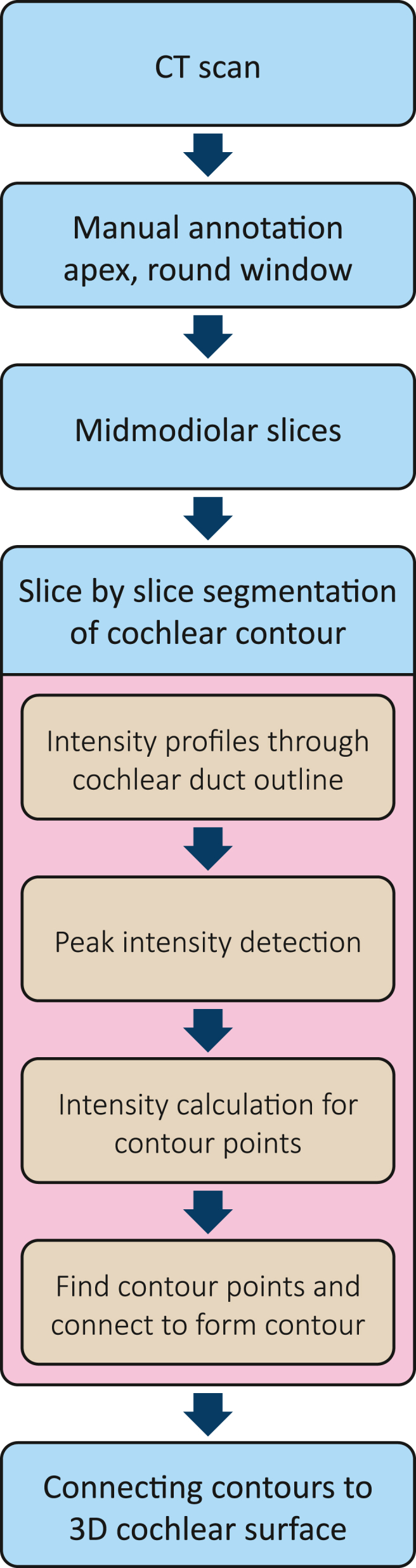
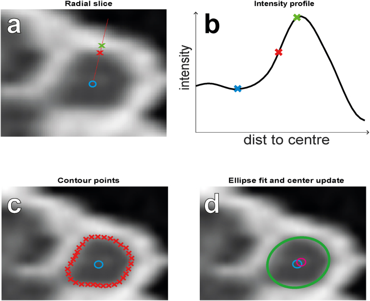
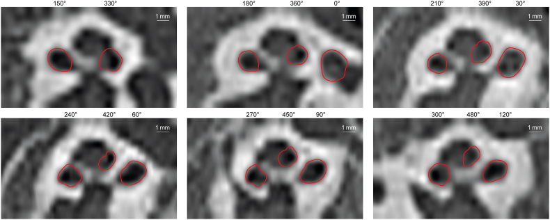
Clinical CT scans were re-oriented and re-sliced to obtain mid-modiolar slices. Using these slices, we segmented the cross-section of the cochlea.

RESULTS

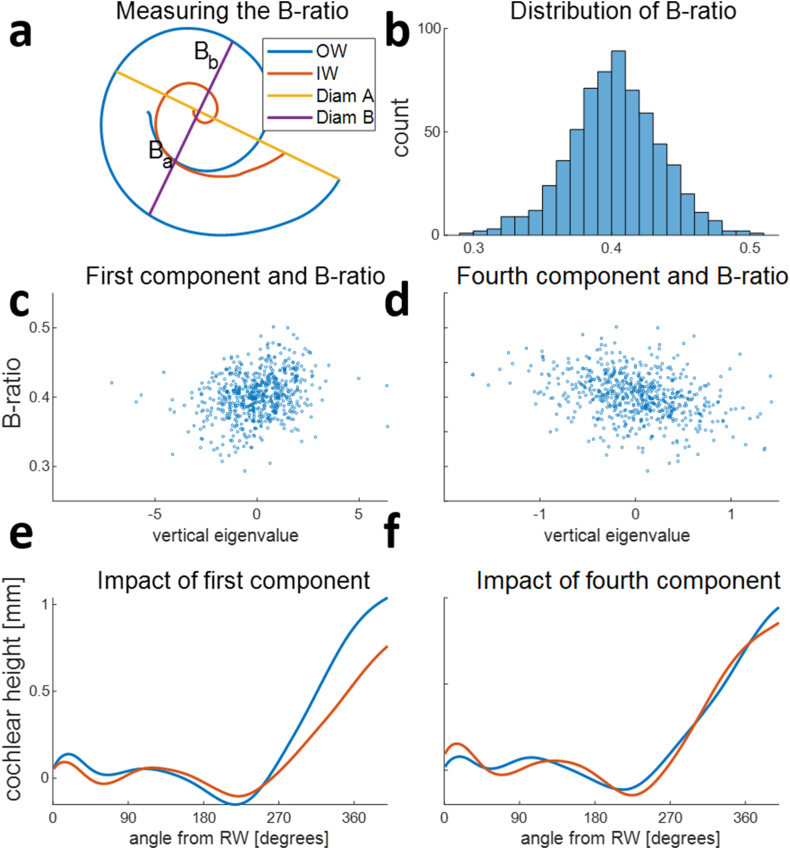
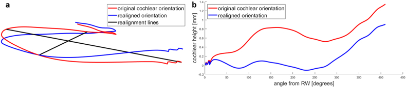
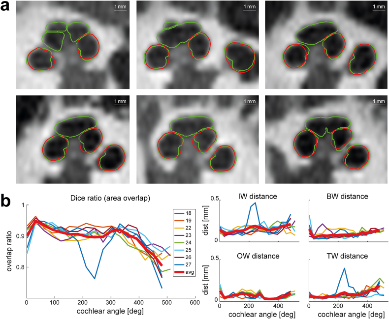
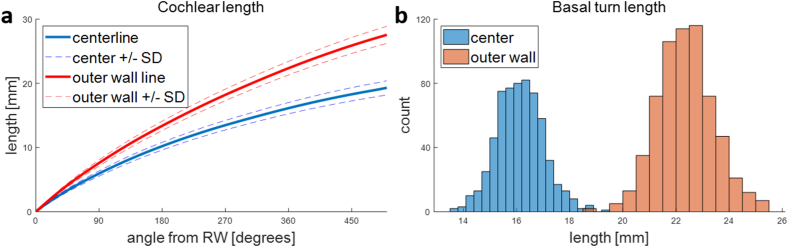
3D surfaces were obtained for the first 1.5 turns of 648 cochleae. Validation of our algorithm against the manually segmented ground truth obtained from 8 micro-CT scans showed good agreement, with 90 % area overlap and an average distance of 0.11 mm between the segmentation contours. The average cochlear duct length for the basal turn was 16.1 mm along the central path and 22.4 mm along the outer wall. The use of an intrinsic, observer-independent coordinate system and principal component analysis enabled unambiguous quantitative evaluation of the vertical trajectory of the cochlea, revealing only a weak correlation between the symmetry of the commonly used basal turn diameters (B-ratio of A and B diameters) and the profile of the vertical trajectory.

CONCLUSION

A model-free segmentation algorithm can achieve similar accuracy as previously published methods relying on statistical shapes. Quantitative analysis of the vertical trajectory can replace the categorization into rollercoaster, sloping, or intermediate vertical trajectory types.

摘要

目的

了解个体患者的耳蜗解剖结构有助于在人工耳蜗植入过程中改进电极选择和放置,以及进行手术规划。本研究的目的是开发一种无模型自动分割算法,从临床计算机断层扫描(CT)中获取描述个体耳蜗解剖结构的三维表面,并可用于定量分析耳蜗的垂直轨迹。

方法

对临床CT扫描进行重新定向和重新切片,以获得中轴切片。利用这些切片,我们对耳蜗的横截面进行了分割。

结果

获得了648个耳蜗前1.5圈的三维表面。将我们的算法与从8次显微CT扫描中手动分割得到的真实情况进行验证,结果显示一致性良好,分割轮廓之间的面积重叠率为90%,平均距离为0.11毫米。基底转的耳蜗管平均长度沿中心路径为16.1毫米,沿外壁为22.4毫米。使用固有、与观察者无关的坐标系和主成分分析能够对耳蜗的垂直轨迹进行明确的定量评估,结果显示常用的基底转直径对称性(A和B直径的B比率)与垂直轨迹轮廓之间仅存在微弱相关性。

结论

无模型分割算法可达到与先前发表的依赖统计形状的方法相似的准确性。对垂直轨迹的定量分析可取代将其分类为过山车型、倾斜型或中间垂直轨迹类型。

https://cdn.ncbi.nlm.nih.gov/pmc/blobs/010f/11367034/42f6352664d3/gr11.jpg
https://cdn.ncbi.nlm.nih.gov/pmc/blobs/010f/11367034/eae4792f6e0b/gr1.jpg
https://cdn.ncbi.nlm.nih.gov/pmc/blobs/010f/11367034/1faa5eba95a7/gr2.jpg
https://cdn.ncbi.nlm.nih.gov/pmc/blobs/010f/11367034/74fa03087f79/gr3.jpg
https://cdn.ncbi.nlm.nih.gov/pmc/blobs/010f/11367034/cc4c0698abc2/gr4.jpg
https://cdn.ncbi.nlm.nih.gov/pmc/blobs/010f/11367034/cbefc75396b6/gr5.jpg
https://cdn.ncbi.nlm.nih.gov/pmc/blobs/010f/11367034/08e42295e3db/gr6.jpg
https://cdn.ncbi.nlm.nih.gov/pmc/blobs/010f/11367034/b97f230356c4/gr7.jpg
https://cdn.ncbi.nlm.nih.gov/pmc/blobs/010f/11367034/a340f9fdcc67/gr8.jpg
https://cdn.ncbi.nlm.nih.gov/pmc/blobs/010f/11367034/d4ee5741b578/gr9.jpg
https://cdn.ncbi.nlm.nih.gov/pmc/blobs/010f/11367034/4a86fc0bd8ce/gr10.jpg
https://cdn.ncbi.nlm.nih.gov/pmc/blobs/010f/11367034/42f6352664d3/gr11.jpg

相似文献

1
Automated segmentation of clinical CT scans of the cochlea and analysis of the cochlea's vertical profile.耳蜗临床CT扫描的自动分割及耳蜗垂直轮廓分析。
Heliyon. 2024 Aug 6;10(16):e35737. doi: 10.1016/j.heliyon.2024.e35737. eCollection 2024 Aug 30.
2
Multi-Scale deep learning framework for cochlea localization, segmentation and analysis on clinical ultra-high-resolution CT images.多尺度深度学习框架在临床超高分辨率 CT 图像上的耳蜗定位、分割和分析。
Comput Methods Programs Biomed. 2020 Jul;191:105387. doi: 10.1016/j.cmpb.2020.105387. Epub 2020 Feb 15.
3
Automatic segmentation of the facial nerve and chorda tympani in pediatric CT scans.小儿 CT 扫描中面神经和鼓索神经的自动分割。
Med Phys. 2011 Oct;38(10):5590-600. doi: 10.1118/1.3634048.
4
Surgical anatomy of the basal turn of the human cochlea as pertaining to cochlear implantation.与人工耳蜗植入相关的人类耳蜗底转的手术解剖学
Otol Neurotol. 2015 Feb;36(2):323-8. doi: 10.1097/MAO.0000000000000371.
5
Atlas-based segmentation of cochlear microstructures in cone beam CT.基于体绘制的锥形束 CT 耳蜗微观结构分割。
Int J Comput Assist Radiol Surg. 2021 Mar;16(3):363-373. doi: 10.1007/s11548-020-02304-x. Epub 2021 Feb 13.
6
Post-operative evaluation of computed tomography imaging following cochlear implantation.人工耳蜗植入术后计算机断层扫描成像的评估
Am J Otolaryngol. 2024 Jan-Feb;45(1):104081. doi: 10.1016/j.amjoto.2023.104081. Epub 2023 Oct 4.
7
Automatic segmentation of intra-cochlear anatomy in post-implantation CT of unilateral cochlear implant recipients.单侧人工耳蜗植入术后 CT 中内耳结构的自动分割。
Med Image Anal. 2014 Apr;18(3):605-15. doi: 10.1016/j.media.2014.02.001. Epub 2014 Feb 18.
8
Imaging of the human cochlea using micro-computed tomography before and after cochlear implantation: comparison with cone-beam computed tomography.使用微计算机断层扫描术在人工耳蜗植入前后对人耳蜗进行成像:与锥形束计算机断层扫描术的比较。
Eur Arch Otorhinolaryngol. 2023 Jul;280(7):3131-3140. doi: 10.1007/s00405-022-07811-y. Epub 2023 Jan 6.
9
Cochlear Implantation: Small Cochlear Diameter May Indicate Degree of Abnormality.人工耳蜗植入术:耳蜗直径小可能表明异常程度。
J Int Adv Otol. 2024 Mar 27;20(2):108-112. doi: 10.5152/iao.2024.231191.
10
Effects of object-to-detector distance and beam energy on synchrotron radiation phase-contrast imaging of implanted cochleae.物距和能束对植入耳蜗的同步辐射相衬成像的影响。
J Microsc. 2019 Feb;273(2):127-134. doi: 10.1111/jmi.12768. Epub 2018 Nov 15.

本文引用的文献

1
Evaluation of a Radiological Tool for Semiautomatic Scalar Translocation Detection After Cochlear Implantation.评估一种用于人工耳蜗植入后半自动化标测移位检测的影像学工具。
Otol Neurotol. 2024 Apr 1;45(4):e322-e327. doi: 10.1097/MAO.0000000000004161. Epub 2024 Feb 20.
2
Automatic electrode scalar location assessment after cochlear implantation using a novel imaging software.使用新型成像软件评估人工耳蜗植入后电极的自动标测位置。
Sci Rep. 2023 Jul 31;13(1):12416. doi: 10.1038/s41598-023-39275-3.
3
Validation of Automatic Cochlear Measurements Using OTOPLAN Software.
使用OTOPLAN软件对耳蜗自动测量进行验证。
J Pers Med. 2023 May 8;13(5):805. doi: 10.3390/jpm13050805.
4
Systematic Literature Review of Hearing Preservation Rates in Cochlear Implantation Associated With Medium- and Longer-Length Flexible Lateral Wall Electrode Arrays.与中长型柔性侧壁电极阵列相关的人工耳蜗植入听力保留率的系统文献综述
Front Surg. 2022 Jul 1;9:893839. doi: 10.3389/fsurg.2022.893839. eCollection 2022.
5
Variation of the cochlear anatomy and cochlea duct length: analysis with a new tablet-based software.耳蜗解剖结构和耳蜗管长度的变异:一种基于平板电脑新软件的分析。
Eur Arch Otorhinolaryngol. 2022 Apr;279(4):1851-1861. doi: 10.1007/s00405-021-06889-0. Epub 2021 May 29.
6
Multi-Scale deep learning framework for cochlea localization, segmentation and analysis on clinical ultra-high-resolution CT images.多尺度深度学习框架在临床超高分辨率 CT 图像上的耳蜗定位、分割和分析。
Comput Methods Programs Biomed. 2020 Jul;191:105387. doi: 10.1016/j.cmpb.2020.105387. Epub 2020 Feb 15.
7
The evaluation of a slim perimodiolar electrode: surgical technique in relation to intracochlear position and cochlear implant outcomes.评价一种纤细的环绕式电极:与耳蜗内位置和人工耳蜗植入效果相关的手术技术。
Eur Arch Otorhinolaryngol. 2020 Feb;277(2):343-350. doi: 10.1007/s00405-019-05696-y. Epub 2019 Oct 24.
8
CT-scan contouring technique allows for direct and reliable measurements of the cochlear duct length: implication in cochlear implantation with straight electrode-arrays.CT 扫描轮廓技术可直接、可靠地测量耳蜗管长度:在直电极阵列人工耳蜗植入中的应用。
Eur Arch Otorhinolaryngol. 2019 Aug;276(8):2135-2140. doi: 10.1007/s00405-019-05432-6. Epub 2019 Apr 22.
9
A Novel Method for Clinical Cochlear Duct Length Estimation toward Patient-Specific Cochlear Implant Selection.一种用于临床耳蜗管长度估计以进行个性化人工耳蜗选择的新方法。
OTO Open. 2018 Oct 2;2(4):2473974X18800238. doi: 10.1177/2473974X18800238. eCollection 2018 Oct-Dec.
10
Challenging aspects of contemporary cochlear implant electrode array design.当代人工耳蜗电极阵列设计的挑战性方面。
World J Otorhinolaryngol Head Neck Surg. 2018 Mar 30;3(4):192-199. doi: 10.1016/j.wjorl.2017.12.007. eCollection 2017 Dec.