• 文献检索
  • 文档翻译
  • 深度研究
  • 学术资讯
  • Suppr Zotero 插件Zotero 插件
  • 邀请有礼
  • 套餐&价格
  • 历史记录
应用&插件
Suppr Zotero 插件Zotero 插件浏览器插件Mac 客户端Windows 客户端微信小程序
定价
高级版会员购买积分包购买API积分包
服务
文献检索文档翻译深度研究API 文档MCP 服务
关于我们
关于 Suppr公司介绍联系我们用户协议隐私条款
关注我们

Suppr 超能文献

核心技术专利:CN118964589B侵权必究
粤ICP备2023148730 号-1Suppr @ 2026

文献检索

告别复杂PubMed语法,用中文像聊天一样搜索,搜遍4000万医学文献。AI智能推荐,让科研检索更轻松。

立即免费搜索

文件翻译

保留排版,准确专业,支持PDF/Word/PPT等文件格式,支持 12+语言互译。

免费翻译文档

深度研究

AI帮你快速写综述,25分钟生成高质量综述,智能提取关键信息,辅助科研写作。

立即免费体验

清除突变的机制驱动乳腺组织的癌变。

Mechanisms that clear mutations drive field cancerization in mammary tissue.

机构信息

VIB-KULeuven Centre for Cancer Biology, Department of Oncology, Leuven, Belgium.

Division of Molecular Pathology, Oncode Institute, The Netherlands Cancer Institute, Amsterdam, the Netherlands.

出版信息

Nature. 2024 Sep;633(8028):198-206. doi: 10.1038/s41586-024-07882-3. Epub 2024 Sep 4.

DOI:10.1038/s41586-024-07882-3
PMID:39232148
原文链接:https://pmc.ncbi.nlm.nih.gov/articles/PMC11374684/
Abstract

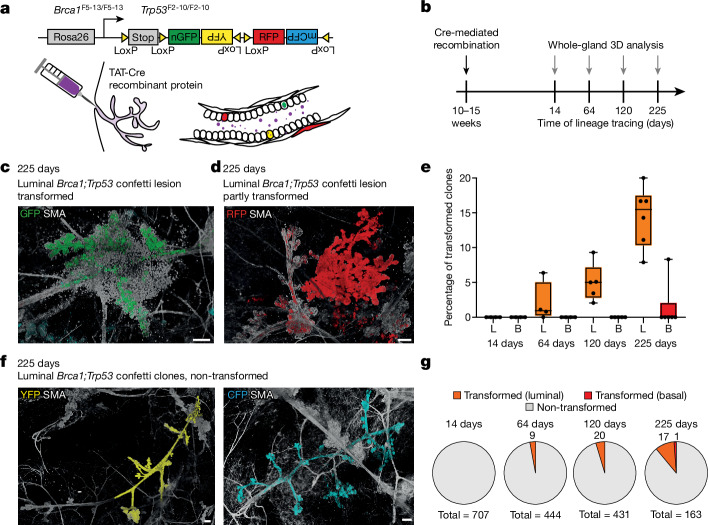
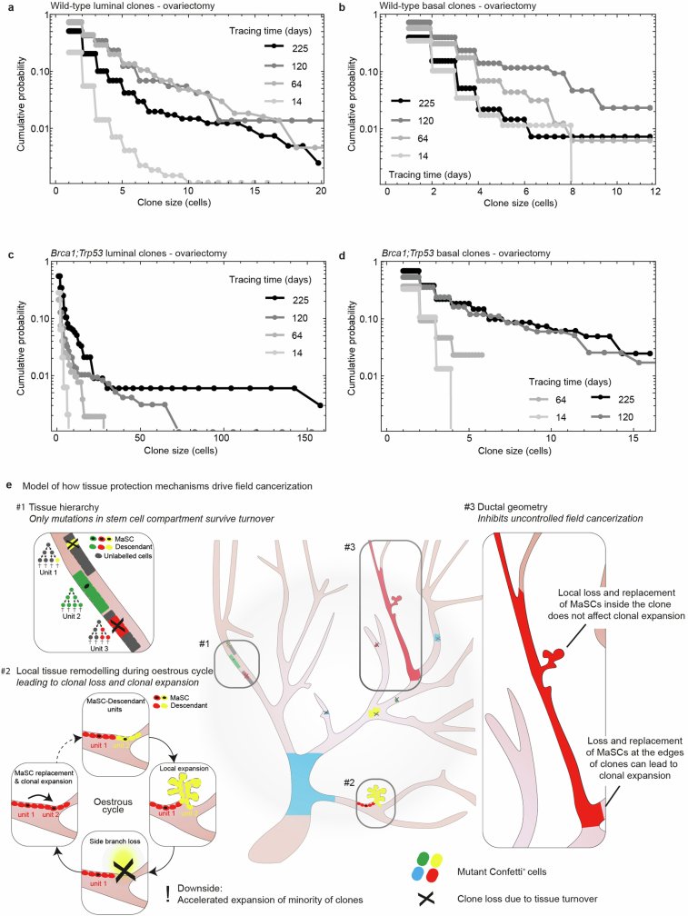
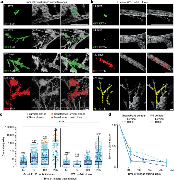
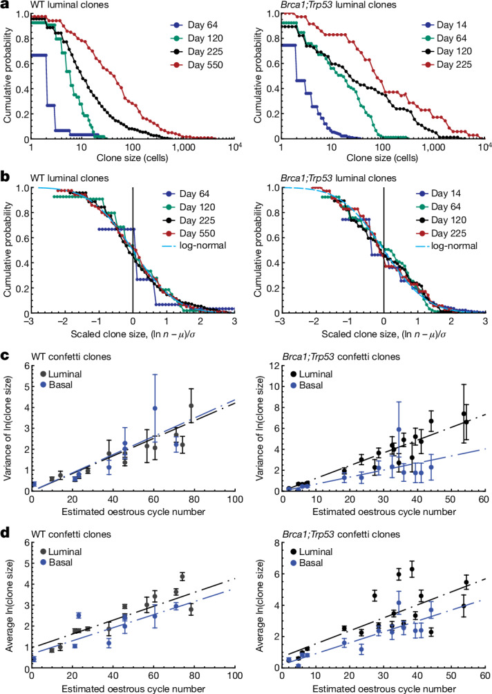
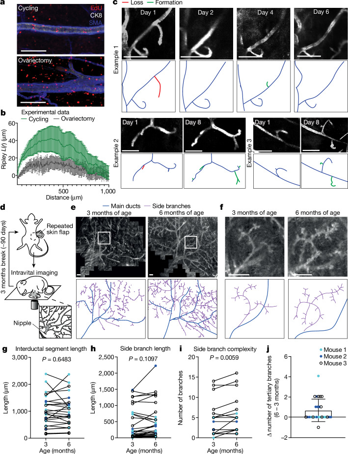
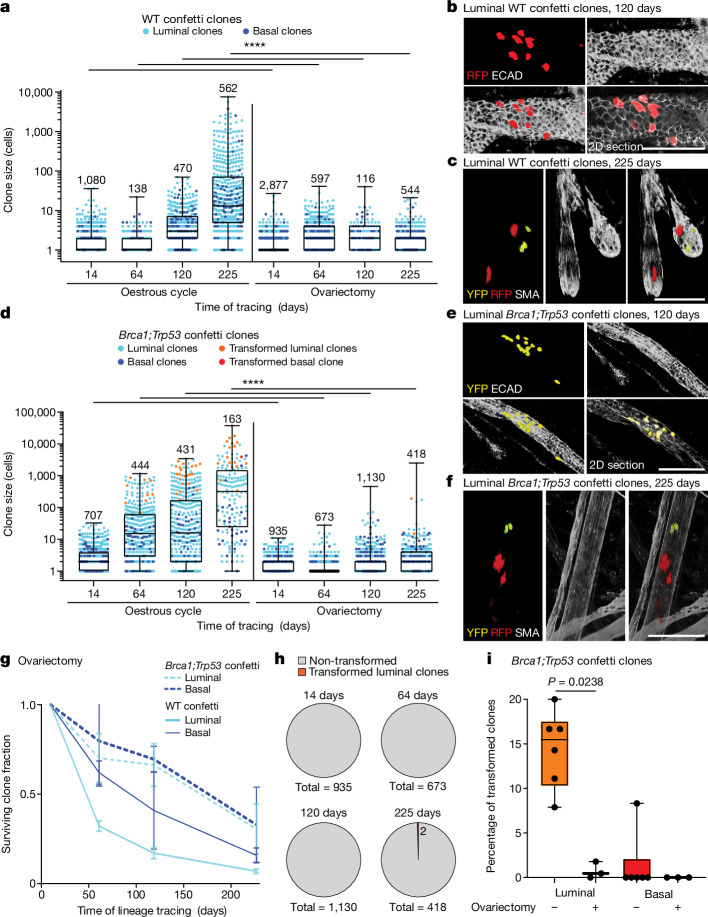
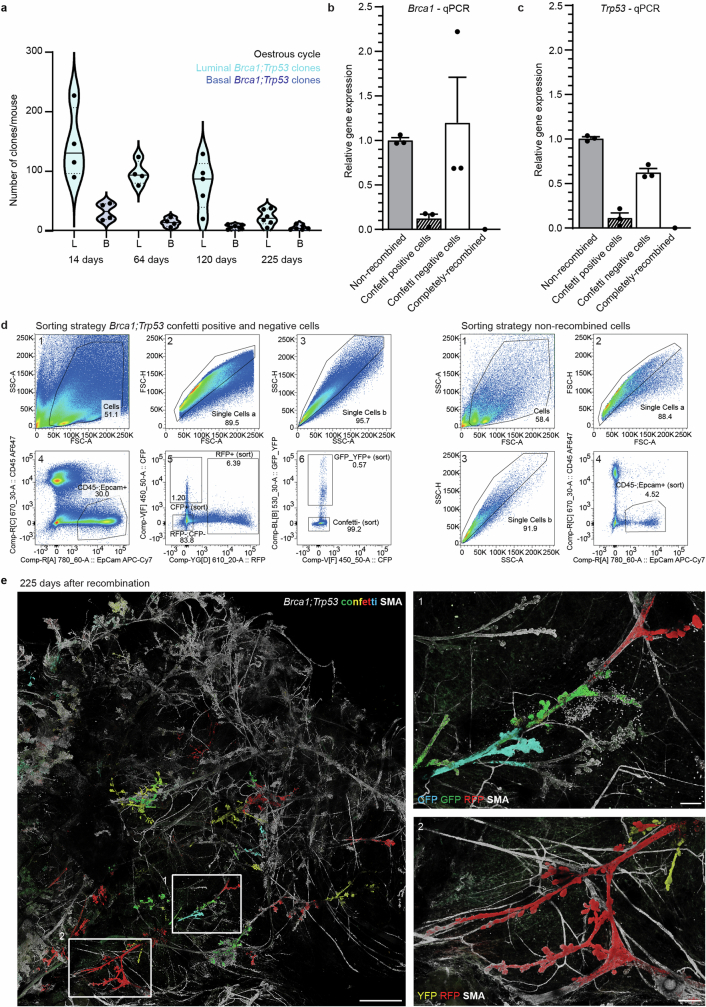
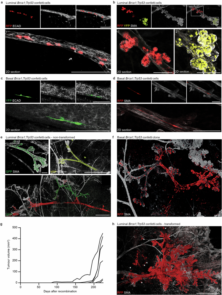
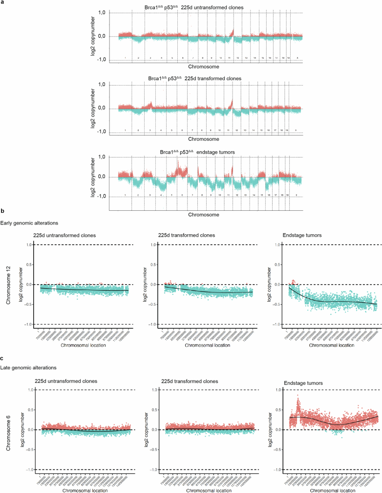
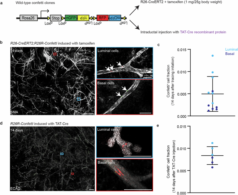
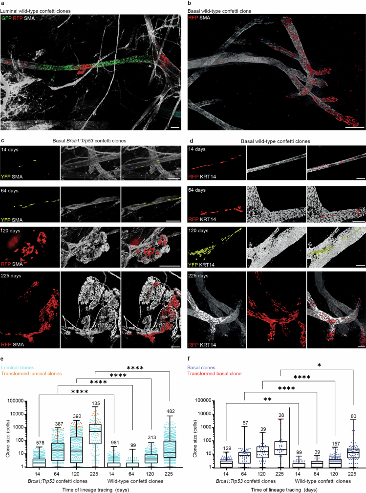
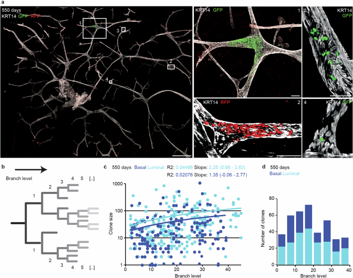
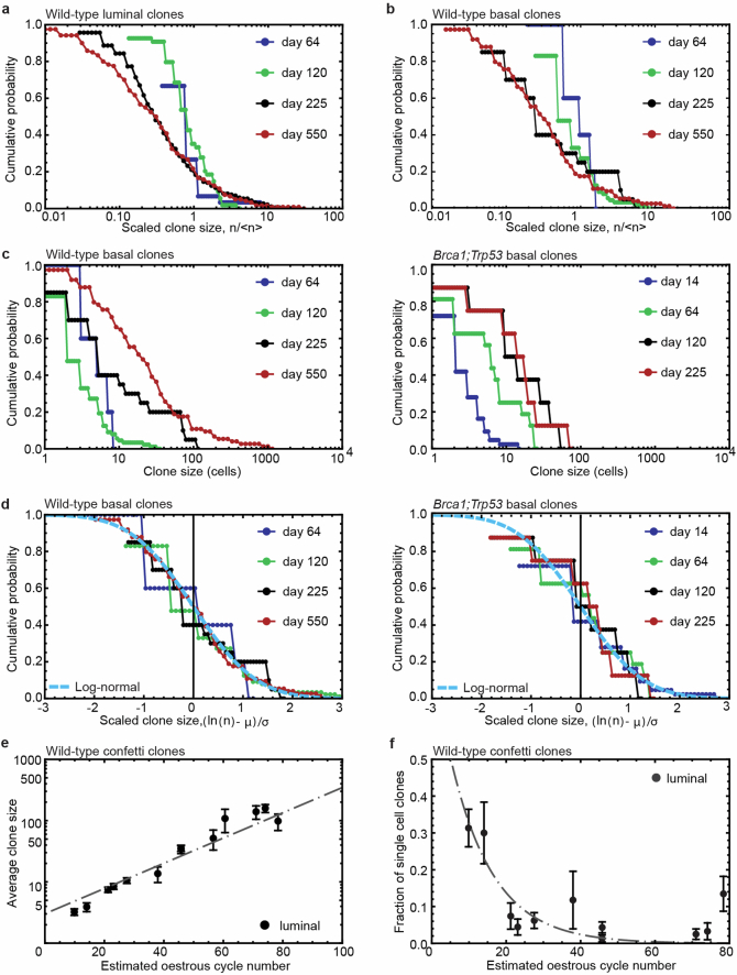
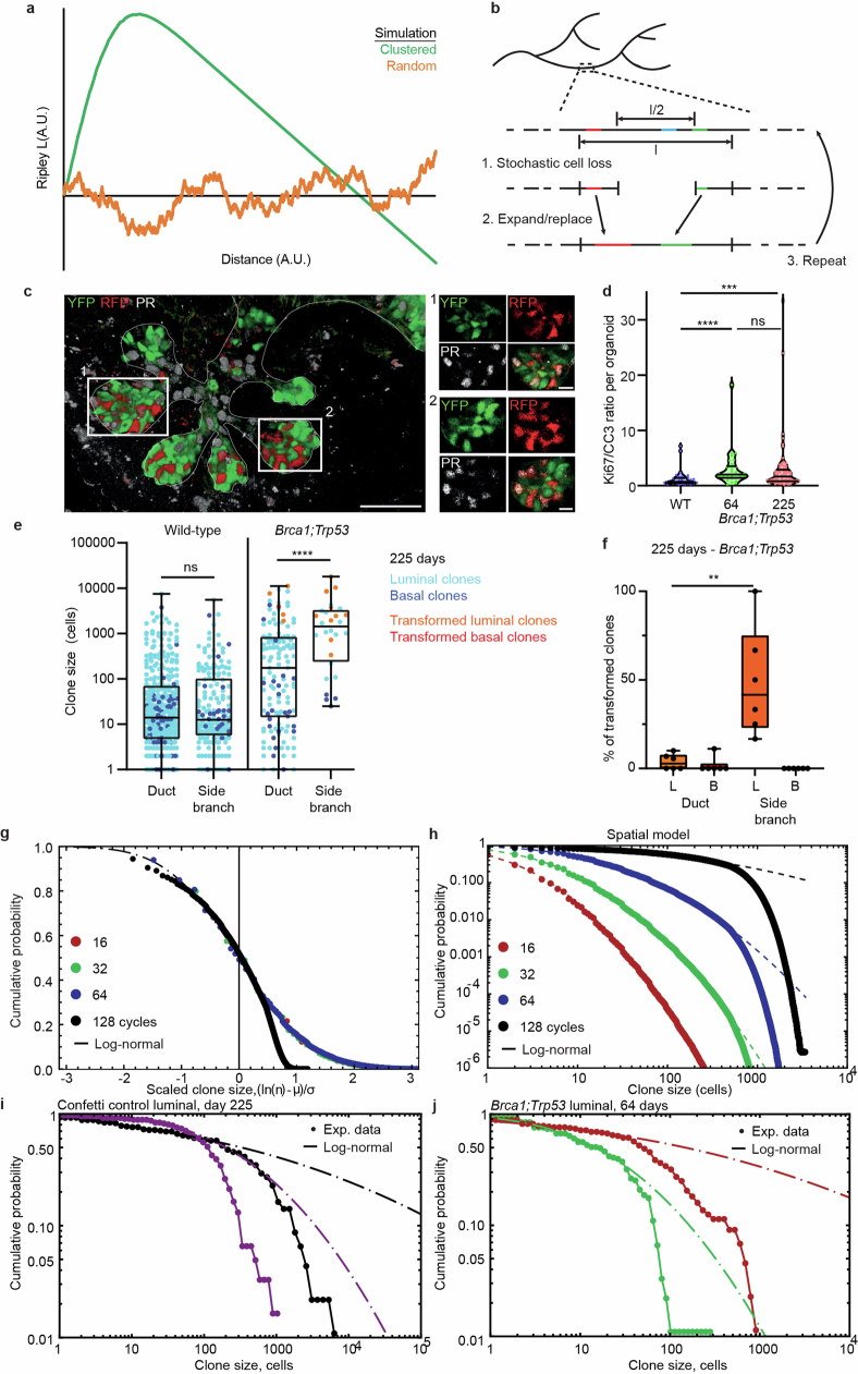
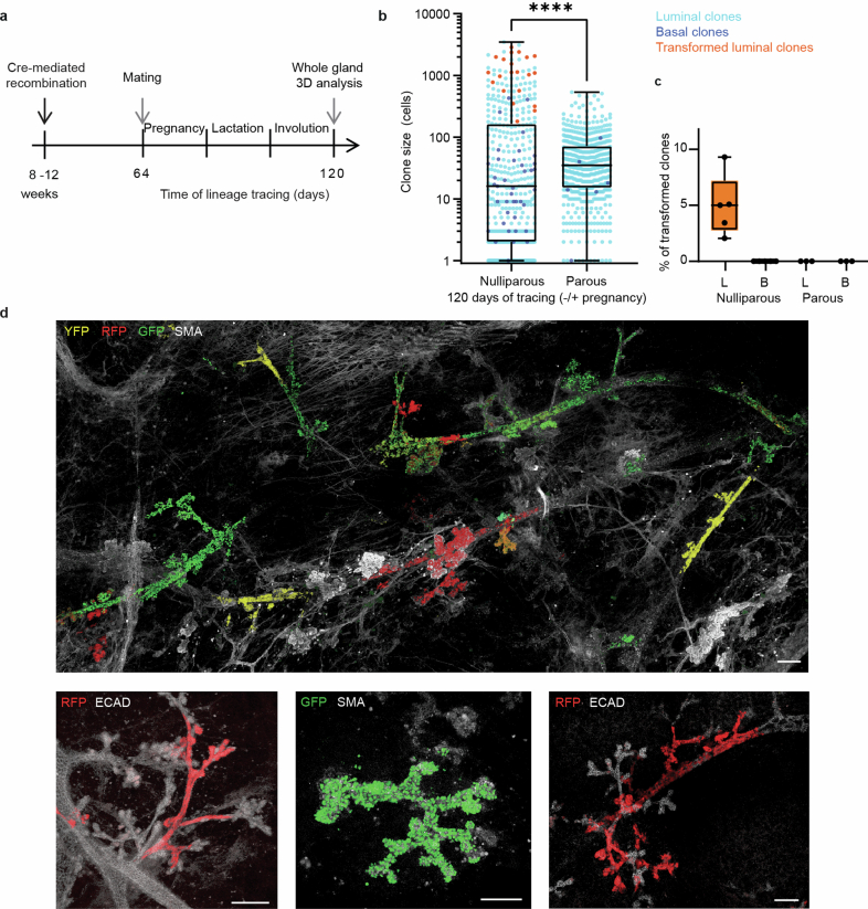
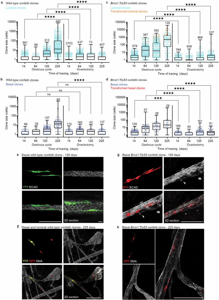
Oncogenic mutations are abundant in the tissues of healthy individuals, but rarely form tumours. Yet, the underlying protection mechanisms are largely unknown. To resolve these mechanisms in mouse mammary tissue, we use lineage tracing to map the fate of wild-type and Brca1;Trp53 cells, and find that both follow a similar pattern of loss and spread within ducts. Clonal analysis reveals that ducts consist of small repetitive units of self-renewing cells that give rise to short-lived descendants. This offers a first layer of protection as any descendants, including oncogenic mutant cells, are constantly lost, thereby limiting the spread of mutations to a single stem cell-descendant unit. Local tissue remodelling during consecutive oestrous cycles leads to the cooperative and stochastic loss and replacement of self-renewing cells. This process provides a second layer of protection, leading to the elimination of most mutant clones while enabling the minority that by chance survive to expand beyond the stem cell-descendant unit. This leads to fields of mutant cells spanning large parts of the epithelial network, predisposing it for transformation. Eventually, clone expansion becomes restrained by the geometry of the ducts, providing a third layer of protection. Together, these mechanisms act to eliminate most cells that acquire somatic mutations at the expense of driving the accelerated expansion of a minority of cells, which can colonize large areas, leading to field cancerization.

摘要

致癌突变在健康个体的组织中很常见,但很少形成肿瘤。然而,潜在的保护机制在很大程度上尚不清楚。为了在小鼠乳腺组织中解析这些机制,我们使用谱系追踪来追踪野生型和 Brca1;Trp53 细胞的命运,发现它们都遵循类似的导管内丢失和扩散模式。克隆分析表明,导管由自我更新细胞的小重复单元组成,这些细胞产生短暂寿命的后代。这提供了第一层保护,因为任何后代,包括致癌突变细胞,都会不断丢失,从而将突变的传播限制在单个干细胞-后代单元内。在连续发情周期中局部组织重塑导致自我更新细胞的协同和随机丢失和替换。这个过程提供了第二层保护,导致大多数突变克隆被消除,同时使少数有机会存活下来的克隆能够扩展到干细胞-后代单元之外。这导致跨越上皮网络大部分区域的突变细胞区域,使其容易发生转化。最终,由于导管的几何形状,克隆扩展受到限制,从而提供了第三层保护。这些机制共同作用,以牺牲驱动少数细胞加速扩张为代价,消除大多数获得体细胞突变的细胞,这些细胞可以殖民大面积,导致癌前病变。

https://cdn.ncbi.nlm.nih.gov/pmc/blobs/cc25/11374684/c6fbd87af00d/41586_2024_7882_Fig17_ESM.jpg
https://cdn.ncbi.nlm.nih.gov/pmc/blobs/cc25/11374684/c13ed1542188/41586_2024_7882_Fig1_HTML.jpg
https://cdn.ncbi.nlm.nih.gov/pmc/blobs/cc25/11374684/c02cdce42216/41586_2024_7882_Fig2_HTML.jpg
https://cdn.ncbi.nlm.nih.gov/pmc/blobs/cc25/11374684/dec788daf982/41586_2024_7882_Fig3_HTML.jpg
https://cdn.ncbi.nlm.nih.gov/pmc/blobs/cc25/11374684/8a90dcfacca2/41586_2024_7882_Fig4_HTML.jpg
https://cdn.ncbi.nlm.nih.gov/pmc/blobs/cc25/11374684/2062f4569df5/41586_2024_7882_Fig5_HTML.jpg
https://cdn.ncbi.nlm.nih.gov/pmc/blobs/cc25/11374684/5871c0615d6e/41586_2024_7882_Fig6_ESM.jpg
https://cdn.ncbi.nlm.nih.gov/pmc/blobs/cc25/11374684/85e2c02a55fc/41586_2024_7882_Fig7_ESM.jpg
https://cdn.ncbi.nlm.nih.gov/pmc/blobs/cc25/11374684/0410b05bfc9f/41586_2024_7882_Fig8_ESM.jpg
https://cdn.ncbi.nlm.nih.gov/pmc/blobs/cc25/11374684/8add89856d86/41586_2024_7882_Fig9_ESM.jpg
https://cdn.ncbi.nlm.nih.gov/pmc/blobs/cc25/11374684/ed6d3460e828/41586_2024_7882_Fig10_ESM.jpg
https://cdn.ncbi.nlm.nih.gov/pmc/blobs/cc25/11374684/99d961acfe97/41586_2024_7882_Fig11_ESM.jpg
https://cdn.ncbi.nlm.nih.gov/pmc/blobs/cc25/11374684/3d74fc0e2620/41586_2024_7882_Fig12_ESM.jpg
https://cdn.ncbi.nlm.nih.gov/pmc/blobs/cc25/11374684/5ce0f0cc94c7/41586_2024_7882_Fig13_ESM.jpg
https://cdn.ncbi.nlm.nih.gov/pmc/blobs/cc25/11374684/2fc17b21b7fe/41586_2024_7882_Fig14_ESM.jpg
https://cdn.ncbi.nlm.nih.gov/pmc/blobs/cc25/11374684/073765f3c4a8/41586_2024_7882_Fig15_ESM.jpg
https://cdn.ncbi.nlm.nih.gov/pmc/blobs/cc25/11374684/8a12590ad7e9/41586_2024_7882_Fig16_ESM.jpg
https://cdn.ncbi.nlm.nih.gov/pmc/blobs/cc25/11374684/c6fbd87af00d/41586_2024_7882_Fig17_ESM.jpg

相似文献

1
Mechanisms that clear mutations drive field cancerization in mammary tissue.清除突变的机制驱动乳腺组织的癌变。
Nature. 2024 Sep;633(8028):198-206. doi: 10.1038/s41586-024-07882-3. Epub 2024 Sep 4.
2
Somatic loss of p53 leads to stem/progenitor cell amplification in both mammary epithelial compartments, basal and luminal.p53 基因的体细胞缺失会导致乳腺上皮细胞的干细胞/祖细胞在基底细胞和腔细胞两个部位扩增。
Stem Cells. 2013 Sep;31(9):1857-67. doi: 10.1002/stem.1429.
3
Alpha-6 integrin deletion delays the formation of Brca1/p53-deficient basal-like breast tumors by restricting luminal progenitor cell expansion.α6 整合素缺失通过限制腔前体细胞扩增延迟了 Brca1/p53 缺陷型基底样乳腺肿瘤的形成。
Breast Cancer Res. 2024 Jun 4;26(1):91. doi: 10.1186/s13058-024-01851-4.
4
Epidermal Tissue Adapts to Restrain Progenitors Carrying Clonal p53 Mutations.表皮组织适应抑制携带克隆性 p53 突变的祖细胞。
Cell Stem Cell. 2018 Nov 1;23(5):687-699.e8. doi: 10.1016/j.stem.2018.08.017. Epub 2018 Sep 27.
5
Atypical cell cycle regulation promotes mammary stem cell expansion during mammary development and tumourigenesis.非典型细胞周期调控促进乳腺发育和肿瘤发生过程中乳腺干细胞的扩增。
Breast Cancer Res. 2024 Jun 28;26(1):106. doi: 10.1186/s13058-024-01862-1.
6
Growth retardation, DNA repair defects, and lack of spermatogenesis in BRCA1-deficient mice.BRCA1基因缺陷小鼠出现生长迟缓、DNA修复缺陷以及精子发生障碍。
Mol Cell Biol. 1999 Oct;19(10):7061-75. doi: 10.1128/MCB.19.10.7061.
7
Time-resolved single-cell analysis of Brca1 associated mammary tumourigenesis reveals aberrant differentiation of luminal progenitors.Brca1 相关乳腺肿瘤发生的时间分辨单细胞分析显示管腔祖细胞的异常分化。
Nat Commun. 2021 Mar 9;12(1):1502. doi: 10.1038/s41467-021-21783-3.
8
Humanization of the mouse mammary gland by replacement of the luminal layer with genetically engineered preneoplastic human cells.通过用基因工程改造的癌前人类细胞替代管腔层来实现小鼠乳腺的人源化。
Breast Cancer Res. 2014 Dec 20;16(6):504. doi: 10.1186/s13058-014-0504-9.
9
c-Kit is required for growth and survival of the cells of origin of Brca1-mutation-associated breast cancer.c-Kit 对于 BRCA1 突变相关乳腺癌的起源细胞的生长和存活是必需的。
Oncogene. 2012 Feb 16;31(7):869-83. doi: 10.1038/onc.2011.289. Epub 2011 Jul 18.
10
The gain of function of p53 cancer mutant in promoting mammary tumorigenesis.p53 癌突变促进乳腺肿瘤发生的功能获得。
Oncogene. 2013 Jun 6;32(23):2900-6. doi: 10.1038/onc.2012.299. Epub 2012 Jul 23.

引用本文的文献

1
Unravelling the genetics and epigenetics of the ageing tumour microenvironment in cancer.解析癌症中衰老肿瘤微环境的遗传学和表观遗传学。
Nat Rev Cancer. 2025 Sep 8. doi: 10.1038/s41568-025-00868-x.
2
The Warburg hypothesis and the emergence of the mitochondrial metabolic theory of cancer.瓦伯格假说与癌症线粒体代谢理论的出现。
J Bioenerg Biomembr. 2025 Apr 8. doi: 10.1007/s10863-025-10059-w.
3
Environmental Drivers of Malignant Clonal Selection in Precancerous Lesion Evolution.癌前病变演变过程中恶性克隆选择的环境驱动因素

本文引用的文献

1
Ductal carcinoma in situ develops within clonal fields of mutant cells in morphologically normal ducts.导管原位癌发生于形态正常的导管中克隆性突变细胞的领域内。
J Pathol. 2024 Jul;263(3):360-371. doi: 10.1002/path.6289. Epub 2024 May 23.
2
Hallmarks of stemness in mammalian tissues.哺乳动物组织中的干性特征。
Cell Stem Cell. 2024 Jan 4;31(1):7-24. doi: 10.1016/j.stem.2023.12.006.
3
The mutational landscape of the adult healthy parous and nulliparous human breast.成年有产和无产健康人类乳房的突变景观。
Cell Prolif. 2025 May;58(5):e70011. doi: 10.1111/cpr.70011. Epub 2025 Feb 25.
4
Oncogene activated human breast luminal progenitors contribute basally located myoepithelial cells.致癌基因激活的人乳腺腔面祖细胞可产生位于基底的肌上皮细胞。
Breast Cancer Res. 2024 Dec 18;26(1):183. doi: 10.1186/s13058-024-01939-x.
5
Estimating the Number of Polygenic Diseases Among Six Mutually Exclusive Entities of Non-Tumors and Cancer.估计六种非肿瘤和癌症相互排斥实体中的多基因疾病数量。
Int J Mol Sci. 2024 Nov 7;25(22):11968. doi: 10.3390/ijms252211968.
6
15th Annual ENBDC Meeting: How do Cellular Potency, Microenvironment and Natural Rhythms Influence Mammary Gland Biology and Breast Cancer?第十五届年度 ENBDC 会议:细胞多能性、微环境和自然节律如何影响乳腺生物学和乳腺癌?
J Mammary Gland Biol Neoplasia. 2024 Nov 25;29(1):19. doi: 10.1007/s10911-024-09572-2.
7
A roadmap to reduce the incidence and mortality of breast cancer by rethinking our approach to women's health.通过重新思考我们对待女性健康的方式来降低乳腺癌发病率和死亡率的路线图。
Breast Cancer Res Treat. 2025 Jan;209(1):1-14. doi: 10.1007/s10549-024-07522-4. Epub 2024 Nov 12.
8
Protection against tumour formation.预防肿瘤形成。
Nat Rev Cancer. 2024 Nov;24(11):741. doi: 10.1038/s41568-024-00762-y.
Nat Commun. 2023 Sep 6;14(1):5136. doi: 10.1038/s41467-023-40608-z.
4
High prevalence of somatic PIK3CA and TP53 pathogenic variants in the normal mammary gland tissue of sporadic breast cancer patients revealed by duplex sequencing.双重测序揭示散发性乳腺癌患者正常乳腺组织中体细胞PIK3CA和TP53致病变异的高发生率。
NPJ Breast Cancer. 2022 Jun 29;8(1):76. doi: 10.1038/s41523-022-00443-9.
5
Breast cancers originating from the terminal ductal lobular units: In situ and invasive acinar adenocarcinoma of the breast, AAB.来源于终末导管小叶单位的乳腺癌:乳腺原位和浸润性腺样囊性癌,AAB。
Eur J Radiol. 2022 Jul;152:110323. doi: 10.1016/j.ejrad.2022.110323. Epub 2022 May 10.
6
Longitudinal Intravital Microscopy Using a Mammary Imaging Window with Replaceable Lid.使用带有可更换盖子的乳腺成像窗口进行纵向活体显微镜检查。
J Vis Exp. 2022 Jan 20(179). doi: 10.3791/63326.
7
Single cell transcriptome atlas of mouse mammary epithelial cells across development.小鼠乳腺上皮细胞发育过程中的单细胞转录组图谱
Breast Cancer Res. 2021 Jun 29;23(1):69. doi: 10.1186/s13058-021-01445-4.
8
An Intravital Microscopy Toolbox to Study Mammary Gland Dynamics from Cellular Level to Organ Scale.一种用于从细胞水平到器官尺度研究乳腺动态的活体显微镜工具包。
J Mammary Gland Biol Neoplasia. 2021 Mar;26(1):9-27. doi: 10.1007/s10911-021-09487-2. Epub 2021 May 4.
9
How should we define mammary stem cells?我们应该如何定义乳腺干细胞?
Trends Cell Biol. 2021 Aug;31(8):621-627. doi: 10.1016/j.tcb.2021.03.012. Epub 2021 Apr 23.
10
Antigen retrieval and clearing for whole-organ immunofluorescence by FLASH.FLASH 进行全器官免疫荧光的抗原修复和通透。
Nat Protoc. 2021 Jan;16(1):239-262. doi: 10.1038/s41596-020-00414-z. Epub 2020 Nov 27.