Figini Matteo, Lin Hongxiang, D'Arco Felice, Ogbole Godwin, Rossi-Espagnet Maria Camilla, Oyinloye Olalekan Ibukun, Yaria Joseph, Nzeh Donald Amasike, Atalabi Mojisola Omolola, Carmichael David W, Cross Judith Helen, Lagunju Ikeoluwa, Fernandez-Reyes Delmiro, Alexander Daniel C
Centre for Medical Image Computing, University College London, 90 High Holborn, London, WC1V 6LJ, UK.
Computer Science, University College London, London, UK.
Neuroradiology. 2024 Dec;66(12):2243-2252. doi: 10.1007/s00234-024-03448-2. Epub 2024 Sep 6.
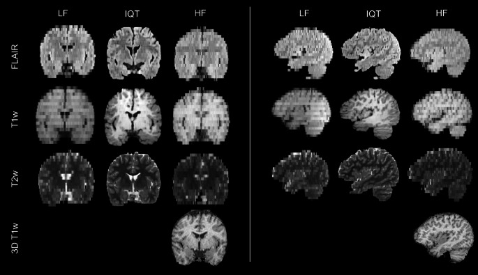
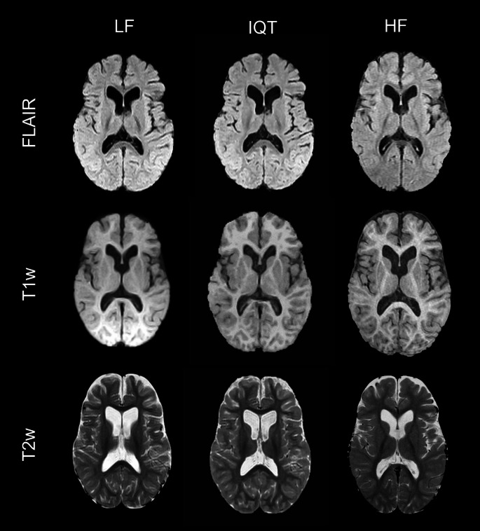
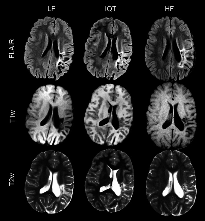
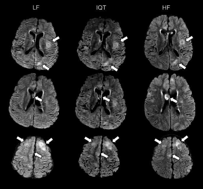
Low-field (LF) MRI scanners are common in many Low- and middle-Income countries, but they provide images with worse spatial resolution and contrast than high-field (HF) scanners. Image Quality Transfer (IQT) is a machine learning framework to enhance images based on high-quality references that has recently adapted to LF MRI. In this study we aim to assess if it can improve lesion visualisation compared to LF MRI scans in children with epilepsy.
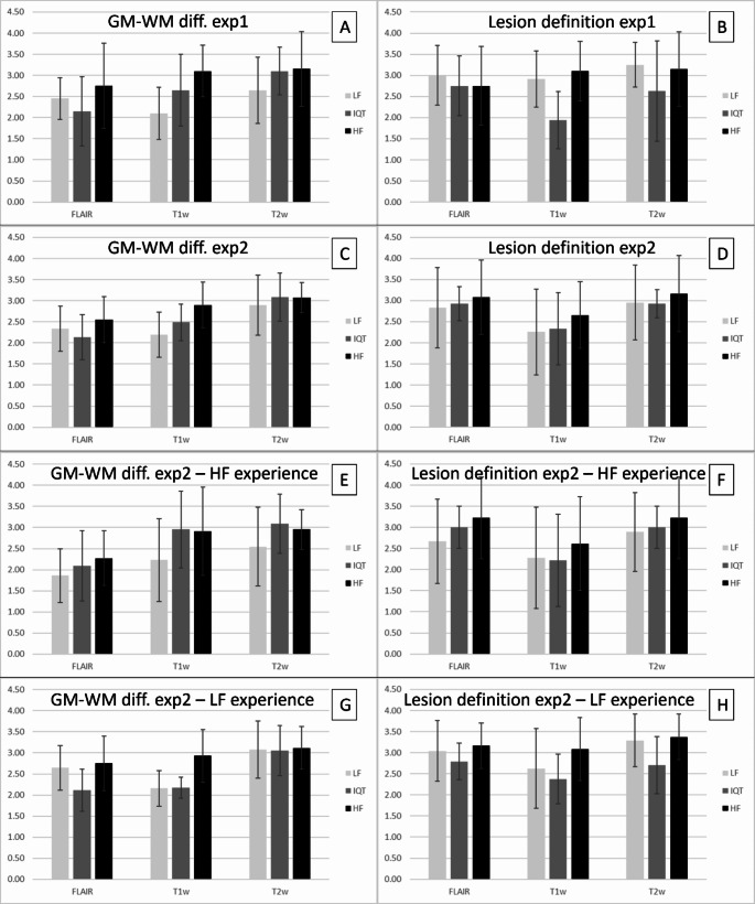
T1-weighted, T2-weighted and FLAIR were acquired from 12 patients (5 to 18 years old, 7 males) with clinical diagnosis of intractable epilepsy on a 0.36T (LF) and a 1.5T scanner (HF). LF images were enhanced with IQT. Seven radiologists blindly evaluated the differentiation between normal grey matter (GM) and white matter (WM) and the extension and definition of epileptogenic lesions in LF, HF and IQT-enhanced images.
When images were evaluated independently, GM-WM differentiation scores of IQT outputs were 26% higher, 17% higher and 12% lower than LF for T1, T2 and FLAIR. Lesion definition scores were 8-34% lower than LF, but became 3% higher than LF for FLAIR and T1 when images were seen side by side. Radiologists with expertise at HF scored IQT images higher than those with expertise at LF.
IQT generally improved the image quality assessments. Evaluation of pathology on IQT-enhanced images was affected by familiarity with HF/IQT image appearance. These preliminary results show that IQT could have an important impact on neuroradiology practice where HF MRI is not available.
低场(LF)磁共振成像(MRI)扫描仪在许多低收入和中等收入国家很常见,但与高场(HF)扫描仪相比,它们提供的图像空间分辨率和对比度更差。图像质量转移(IQT)是一种基于高质量参考图像来增强图像的机器学习框架,最近已应用于LF MRI。在本研究中,我们旨在评估与癫痫患儿的LF MRI扫描相比,IQT是否能改善病变的可视化。
对12例临床诊断为难治性癫痫的患者(年龄5至18岁,男性7例)在0.36T(LF)和1.5T扫描仪(HF)上采集T1加权、T2加权和液体衰减反转恢复(FLAIR)图像。LF图像用IQT进行增强。7名放射科医生对LF、HF和IQT增强图像中正常灰质(GM)和白质(WM)之间的区分以及致痫性病变的范围和清晰度进行了盲法评估。
独立评估图像时,IQT输出的GM-WM区分分数在T1、T2和FLAIR序列上分别比LF高26%、17%和低12%。病变清晰度分数比LF低8%-34%,但在并排观察图像时,FLAIR和T1序列的分数比LF高3%。具有HF专业知识的放射科医生对IQT图像的评分高于具有LF专业知识者。
IQT总体上改善了图像质量评估。对IQT增强图像上病变的评估受对HF/IQT图像外观熟悉程度的影响。这些初步结果表明,在无法获得HF MRI的情况下,IQT可能会对神经放射学实践产生重要影响。