From the Athinoula A. Martinos Center for Biomedical Imaging (J.E.I., M.S.R.), Department of Radiology (J.E.I., P.S., M.S.R.), Department of Neurology and Center for Genomic Medicine (R.S., W.T.K.), and Department of Emergency Medicine (B.M., J.N.G.), Massachusetts General Hospital and Harvard Medical School, 55 Fruit St, Boston, MA 02114; Centre for Medical Image Computing, Department of Medical Physics and Biomedical Engineering, University College London, London, UK (J.E.I., B.B.); Computer Science and Artificial Intelligence Laboratory, Massachusetts Institute of Technology, Cambridge, Mass (J.E.I.); Swiss Federal Institute of Technology (ETH), Zurich, Switzerland (S.L.); Department of Neurology, Yale New Haven Hospital, New Haven, Conn (K.N.S.); and Department of Physics, Harvard University, Cambridge, Mass (M.S.R.).
Radiology. 2023 Mar;306(3):e220522. doi: 10.1148/radiol.220522. Epub 2022 Nov 8.
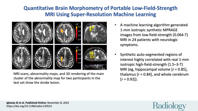
Background Portable, low-field-strength (0.064-T) MRI has the potential to transform neuroimaging but is limited by low spatial resolution and low signal-to-noise ratio. Purpose To implement a machine learning super-resolution algorithm that synthesizes higher spatial resolution images (1-mm isotropic) from lower resolution T1-weighted and T2-weighted portable brain MRI scans, making them amenable to automated quantitative morphometry. Materials and Methods An external high-field-strength MRI data set (1-mm isotropic scans from the Open Access Series of Imaging Studies data set) and segmentations for 39 regions of interest (ROIs) in the brain were used to train a super-resolution convolutional neural network (CNN). Secondary analysis of an internal test set of 24 paired low- and high-field-strength clinical MRI scans in participants with neurologic symptoms was performed. These were part of a prospective observational study (August 2020 to December 2021) at Massachusetts General Hospital (exclusion criteria: inability to lay flat, body habitus preventing low-field-strength MRI, presence of MRI contraindications). Three well-established automated segmentation tools were applied to three sets of scans: high-field-strength (1.5-3 T, reference standard), low-field-strength (0.064 T), and synthetic high-field-strength images generated from the low-field-strength data with the CNN. Statistical significance of correlations was assessed with Student tests. Correlation coefficients were compared with Steiger tests. Results Eleven participants (mean age, 50 years ± 14; seven men) had full cerebrum coverage in the images without motion artifacts or large stroke lesion with distortion from mass effect. Direct segmentation of low-field-strength MRI yielded nonsignificant correlations with volumetric measurements from high field strength for most ROIs ( > .05). Correlations largely improved when segmenting the synthetic images: values were less than .05 for all ROIs (eg, for the hippocampus [ = 0.85; < .001], thalamus [ = 0.84; = .001], and whole cerebrum [ = 0.92; < .001]). Deviations from the model ( score maps) visually correlated with pathologic abnormalities. Conclusion This work demonstrated proof-of-principle augmentation of portable MRI with a machine learning super-resolution algorithm, which yielded highly correlated brain morphometric measurements to real higher resolution images. © RSNA, 2022 See also the editorial by Ertl-Wagner amd Wagner in this issue.
背景便携式、低磁场强度(0.064-T)MRI 有可能改变神经影像学,但受到低空间分辨率和低信噪比的限制。目的 实施机器学习超分辨率算法,从较低分辨率的 T1 加权和 T2 加权便携式脑部 MRI 扫描中合成更高空间分辨率的图像(1 毫米各向同性),使其适用于自动定量形态测量。材料与方法 使用外部高磁场强度 MRI 数据集(来自 Open Access Series of Imaging Studies 数据集的 1 毫米各向同性扫描)和大脑 39 个感兴趣区域(ROI)的分割,训练一个超分辨率卷积神经网络(CNN)。对 24 例有神经症状的参与者的低和高磁场强度临床 MRI 扫描的内部测试集进行二次分析。这些是马萨诸塞州总医院(排除标准:无法平躺、体型阻止低磁场强度 MRI、存在 MRI 禁忌症)一项前瞻性观察研究(2020 年 8 月至 2021 年 12 月)的一部分。应用三种成熟的自动分割工具对三组扫描进行分析:高磁场强度(1.5-3 T,参考标准)、低磁场强度(0.064 T)和从低磁场强度数据用 CNN 生成的合成高磁场强度图像。采用学生 t 检验评估相关性的统计学意义。使用 Steiger 检验比较相关系数。结果 11 名参与者(平均年龄 50 岁±14 岁;7 名男性)的图像无运动伪影且无大卒中病灶,无明显失真。对无运动伪影且无大卒中病灶的低磁场强度 MRI 进行直接分割,与大多数 ROI 的高磁场强度体积测量无显著相关性(>.05)。当对合成图像进行分割时,相关性有很大改善:所有 ROI 的 值均小于.05(例如,海马体 [ = 0.85; <.001]、丘脑 [ = 0.84; =.001] 和整个大脑 [ = 0.92; <.001])。与模型的偏差(得分图)与病理异常明显相关。结论 本研究证明了使用机器学习超分辨率算法增强便携式 MRI 的原理验证,该算法生成的脑形态测量结果与真实的高分辨率图像高度相关。