Bogdańska-Chomczyk E, Wojtacha P, Tsai M L, Huang A C W, Kozłowska A
Department of Human Physiology and Pathophysiology, School of Medicine, Collegium Medicum, University of Warmia and Mazury in Olsztyn, Olsztyn, Poland.
Department of Psychology and Sociology of Health and Public Health, University of Warmia and Mazury in Olsztyn, Olsztyn, Poland.
Front Mol Neurosci. 2024 Aug 23;17:1414457. doi: 10.3389/fnmol.2024.1414457. eCollection 2024.
Attention-deficit/hyperactivity disorder (ADHD) is a neurodevelopmental disorder whose exact pathophysiology has not been fully understood yet. Numerous studies have suggested disruptions in the cellular architecture and neuronal activity within brain structures of individuals with ADHD, accompanied by imbalances in the immune system, oxidative stress, and metabolism.
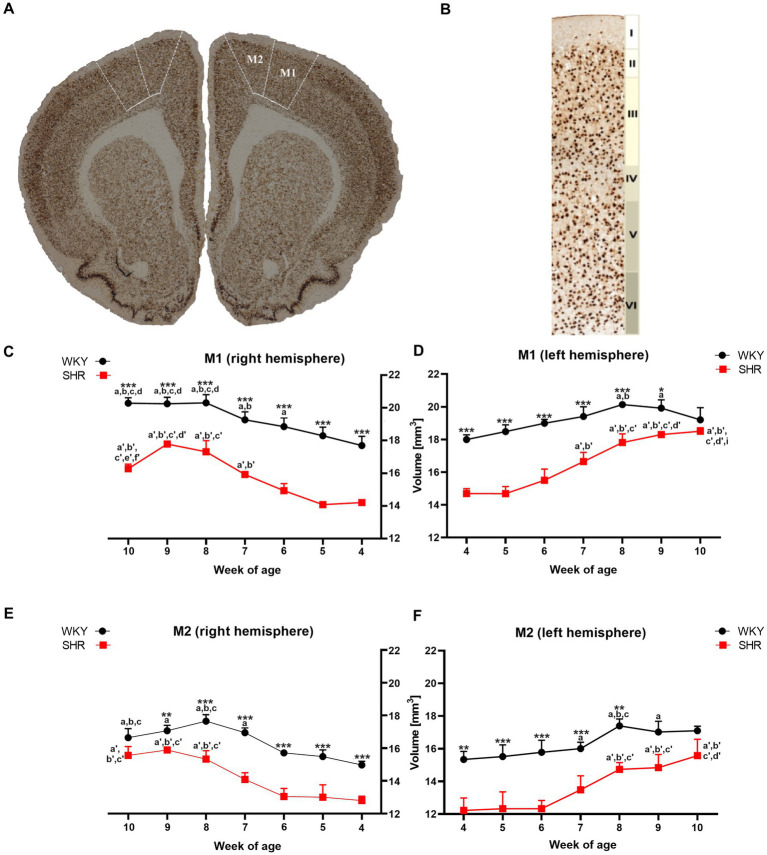
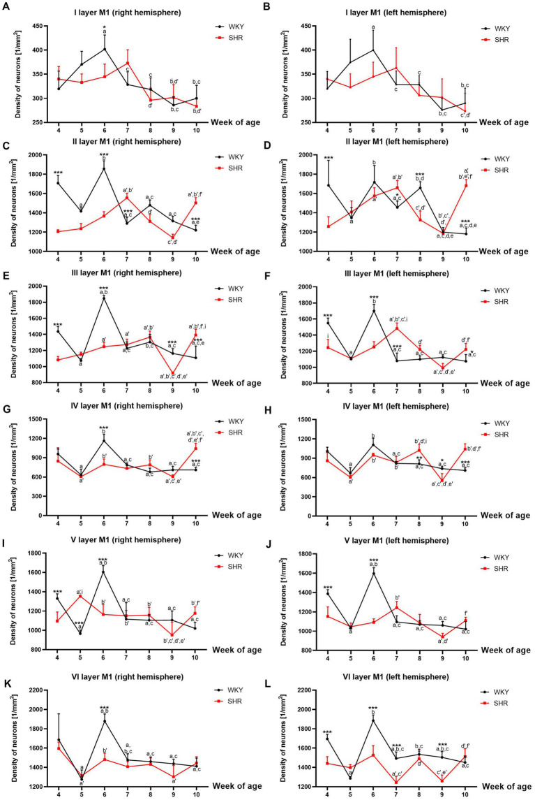
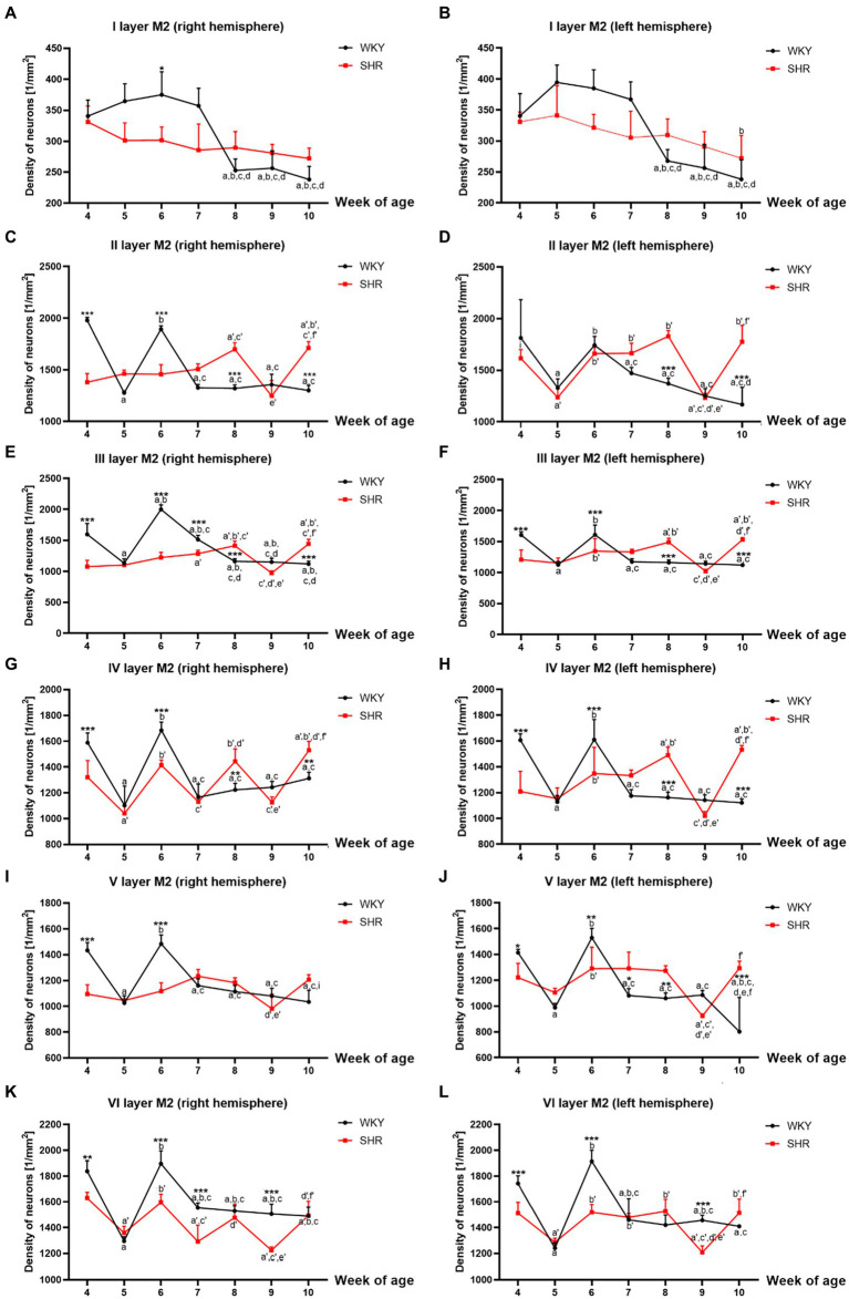
This study aims to assess two functionally and histologically distinct brain areas involved in motor control and coordination: the motor cortex (MC) and prefrontal cortex (PFC). Namely, the morphometric analysis of the MC throughout the developmental stages of Spontaneously Hypertensive Rats (SHRs) and Wistar Kyoto Rats (WKYs). Additionally, the study aimed to investigate the levels and activities of specific immune, oxidative stress, and metabolic markers in the PFC of juvenile and maturing SHRs in comparison to WKYs.
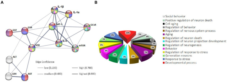
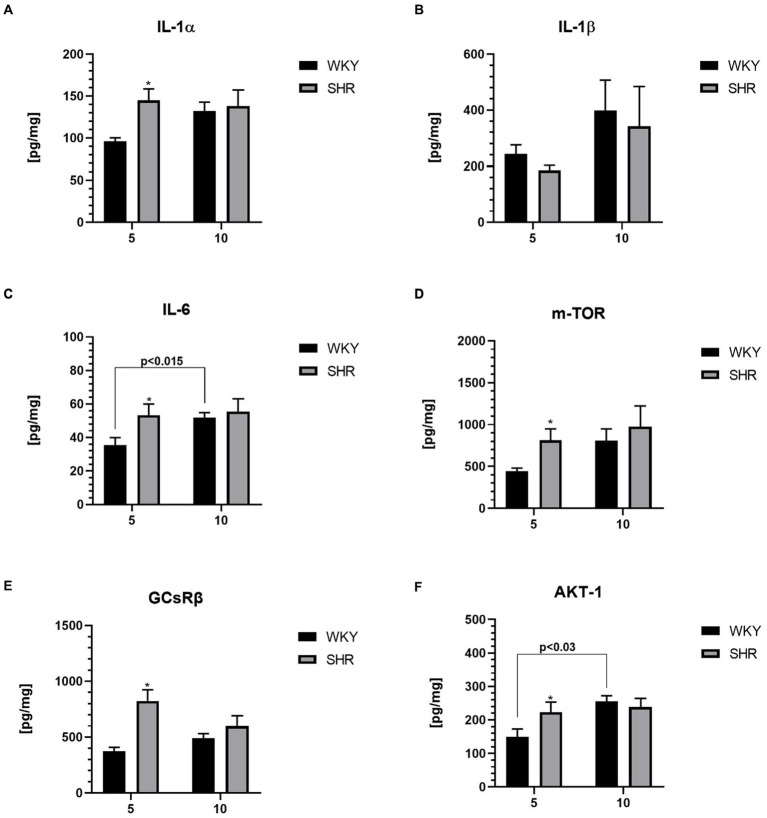
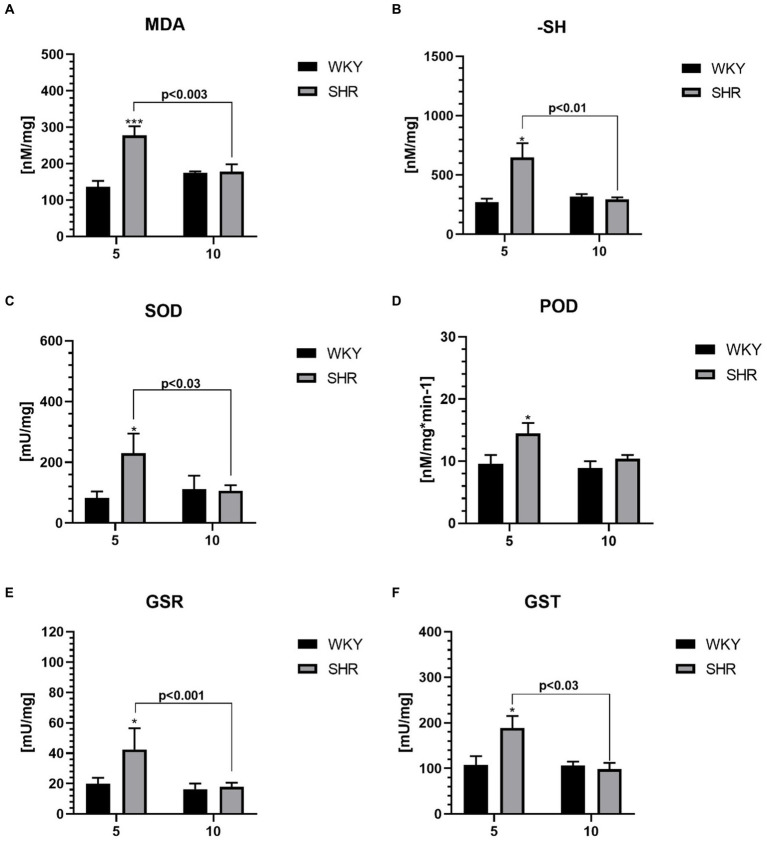
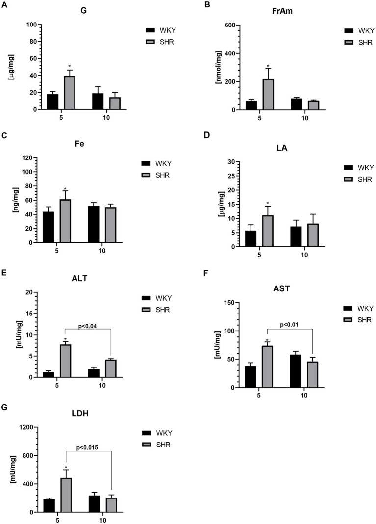
The most significant MC volume reductions occurred in juvenile SHRs, accompanied by alterations in neuronal density in these brain areas compared to WKYs. Furthermore, juvenile SHRs exhibit heightened levels and activity of various markers, including interleukin-1α (IL-1α), IL-6, serine/threonine-protein mammalian target of rapamycin, RAC-alpha serine/threonine-protein kinase, glucocorticoid receptor β, malondialdehyde, sulfhydryl groups, superoxide dismutase, peroxidase, glutathione reductase, glutathione S-transferase, glucose, fructosamine, iron, lactic acid, alanine, aspartate transaminase, and lactate dehydrogenase.
Significant changes in the MC morphometry and elevated levels of inflammatory, oxidative, and metabolic markers in PFC might be associated with disrupted brain development and maturation in ADHD.
注意力缺陷多动障碍(ADHD)是一种神经发育障碍,其确切的病理生理学尚未完全明确。大量研究表明,ADHD患者脑结构中的细胞结构和神经元活动受到破坏,同时伴有免疫系统、氧化应激和代谢失衡。
本研究旨在评估参与运动控制和协调的两个在功能和组织学上不同的脑区:运动皮层(MC)和前额叶皮层(PFC)。具体而言,对自发性高血压大鼠(SHRs)和Wistar Kyoto大鼠(WKYs)发育各阶段的MC进行形态计量分析。此外,该研究旨在比较幼年和成年SHRs与WKYs的PFC中特定免疫、氧化应激和代谢标志物的水平及活性。
幼年SHRs的MC体积减小最为显著,与WKYs相比,这些脑区的神经元密度也发生了改变。此外,幼年SHRs的多种标志物水平和活性升高,包括白细胞介素-1α(IL-1α)、IL-6、雷帕霉素哺乳动物靶蛋白丝氨酸/苏氨酸激酶、RAC-α丝氨酸/苏氨酸蛋白激酶、糖皮质激素受体β、丙二醛、巯基、超氧化物歧化酶、过氧化物酶、谷胱甘肽还原酶、谷胱甘肽S-转移酶、葡萄糖、果糖胺、铁、乳酸、丙氨酸、天冬氨酸转氨酶和乳酸脱氢酶。
MC形态计量的显著变化以及PFC中炎症、氧化和代谢标志物水平的升高可能与ADHD患者大脑发育和成熟的破坏有关。